. 2011 Mar;14(2):205-15.
doi: 10.3109/10253890.2010.531331.

Regulatory interactions of stress and reward on rat forebrain opioidergic and GABAergic circuitry

Affiliations

Regulatory interactions of stress and reward on rat forebrain opioidergic and GABAergic circuitry

A M Christiansen et al. Stress. 2011 Mar.

Abstract

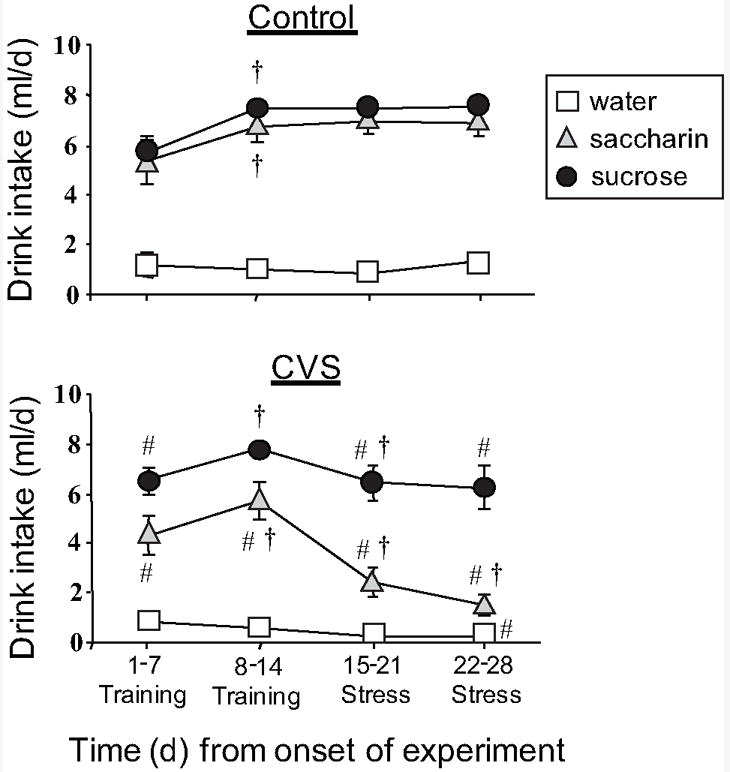
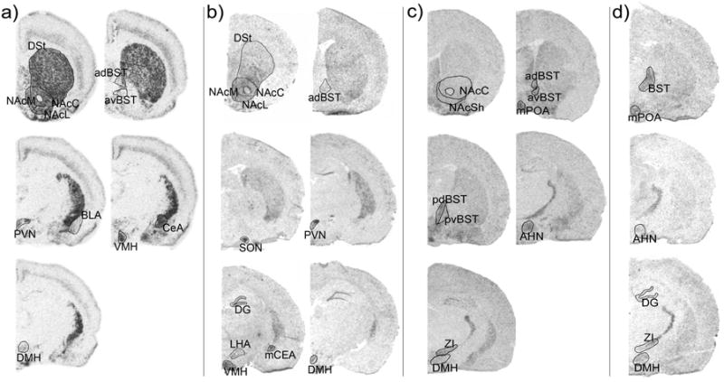
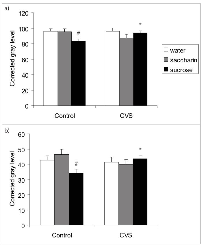
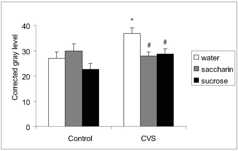
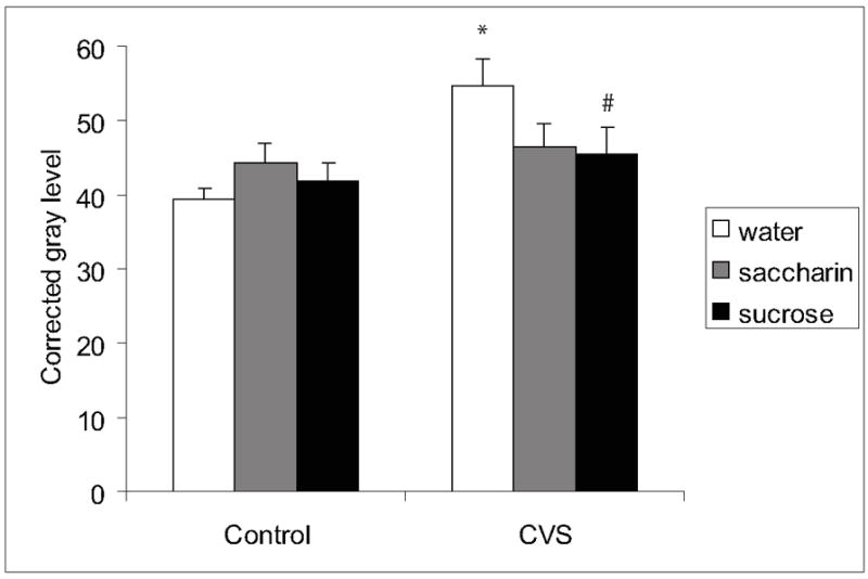
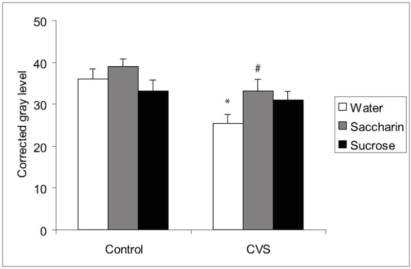
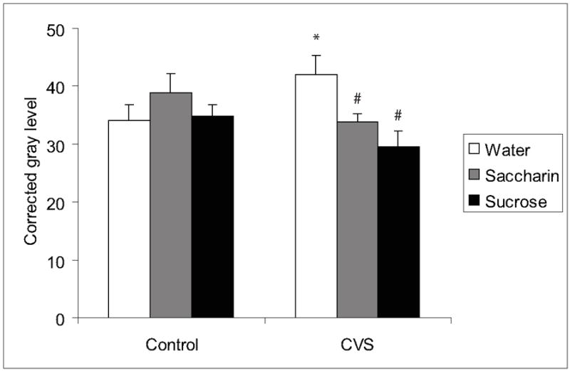
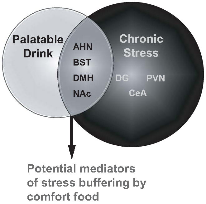
Palatable food intake reduces stress responses, suggesting that individuals may consume such ?comfort? food as self-medication for stress relief. The mechanism by which palatable foods provide stress relief is not known, but likely lies at the intersection of forebrain reward and stress regulatory circuits. Forebrain opioidergic and gamma-aminobutyric acid ergic signaling is critical for both reward and stress regulation, suggesting that these systems are prime candidates for mediating stress relief by palatable foods. Thus, the present study (1) determines how palatable ?comfort? food alters stress-induced changes in the mRNA expression of inhibitory neurotransmitters in reward and stress neurocircuitry and (2) identifies candidate brain regions that may underlie comfort food-mediated stress reduction. We used a model of palatable ?snacking? in combination with a model of chronic variable stress followed by in situ hybridization to determine forebrain levels of pro-opioid and glutamic acid decarboxylase (GAD) mRNA. The data identify regions within the extended amygdala, striatum, and hypothalamus as potential regions for mediating hypothalamic-pituitary-adrenal axis buffering following palatable snacking. Specifically, palatable snacking alone decreased pro-enkephalin-A (ENK) mRNA expression in the anterior bed nucleus of the stria terminalis (BST) and the nucleus accumbens, and decreased GAD65 mRNA in the posterior BST. Chronic stress alone increased ENK mRNA in the hypothalamus, nucleus accumbens, amygdala, and hippocampus; increased dynorphin mRNA in the nucleus accumbens; increased GAD65 mRNA in the anterior hypothalamus and BST; and decreased GAD65 mRNA in the dorsal hypothalamus. Importantly, palatable food intake prevented stress-induced gene expression changes in subregions of the hypothalamus, BST, and nucleus accumbens. Overall, these data suggest that complex interactions exist between brain reward and stress pathways and that palatable snacking can mitigate many of the neurochemical alterations induced by chronic stress.


Conflict of interest statement

DECLARATION OF INTEREST The authors have no conflict of interest. The authors alone are responsible for the content and writing of the paper.

Figures

Figure 1
Figure 1
Timecourse of mean daily drink intake of sucrose, saccharin or water (8ml/day max) in control (a) and CVS (b) rats receiving twice daily access to these drink solutions in addition to ad libitum food and water. Palatable drink intake was reduced during chronic stress. All sucrose and saccharin points were different from water (p<0.05). An # indicates p<0.05 vs. water and a cross indicates p<0.05 vs. the previous timepoint by post-hoc analysis. Abbreviations: chronic variable stress (CVS). Graph reproduced from Ulrich-Lai, et al. 2007, Copyright 2007 The Endocrine Society.
Figure 2
Figure 2
Representative images and regions analyzed for ENK (a), DYN (b), GAD65 (c), and GAD67 (d) mRNA expression by in situ hybridization. Abbreviations: anterior hypothalamic nucleus (AHN), basolateral amygdala (BLA), the bed nucleus of the stria terminalis (BST) (anterodorsal (adBST), anteroventral (avBST), posterior (posteriorBST), posterior dorsal (pdBST), posterior ventral (pvBST), central amygdala (CeA), dentate gyrus (DG), dorsal striatum (DSt), dorsomedial hypothalamic nucleus (DMH), lateral hypothalamic area (LHA), medial amygdala (MeA), medial preoptic area (mPOA), nucleus accumbens NAc (medial shell (NAcM), lateral shell (NAcL), and core (NAcC)), paraventricular hypothalamic nucleus (PVN), ventromedial hypothalamic nucleus (VMH), and zona inserta (ZI). Images for each probe are displayed rostral to caudal from left to right with the most rostral image at the top left.
Figure 3
Figure 3
ENK mRNA in the anterior bed nucleus of the stria terminalis (BST). Sucrose drink reduced ENK mRNA expression in the anterior BST and this reduction was prevented by chronic stress in both the anterodorsal area (a) and the anteroventral area (b). An * indicates a significant difference from non-CVS and a # indicates a significant difference from water by post-hoc analysis. Data are shown as mean ± SEM with n=5-9 per group.
Figure 4
Figure 4
ENK mRNA in the DMH. Chronic stress increased ENK and this increase was prevented by palatable drink in the DMH. An * indicates a significant difference from non-CVS control and a # indicates a significant difference from water by post-hoc analysis. Data are shown as mean ± SEM with n=5-9 per group.
Figure 5
Figure 5
DYN mRNA in the NAc core. Chronic stress increased DYN mRNA expression in the NAc core and this increase was prevented by sucrose or saccharin drink. An * indicates a significant difference from non-CVS control and a # indicates a significant difference from water by post-hoc analysis. Data are shown as mean ± SEM with n=5-9 per group.
Figure 6
Figure 6
GAD65 mRNA in the DMH. Chronic stress decreased GAD65 mRNA in the DMH and this effect was prevented by palatable drink. An * indicates a significant difference from non-CVS control and a # indicates a significant difference from water by post-hoc analysis. Data are shown as mean ± SEM with n=5-9 per group.
Figure 7
Figure 7
GAD65 mRNA in the AHA. Chronic stress increased GAD65 mRNA in the AHA and palatable drink prevented these effects. An * indicates a significant difference from non-CVS control and a # indicates a significant difference from water by post-hoc analysis. Data are shown as mean ± SEM with n=5-9 per group.
Figure 8
Figure 8
Summary diagram of neuroanatomical changes in response to palatable snacking and chronic stress. In a number of brain regions, shown in the overlapping circles, neurochemical changes induced by chronic variable stress were prevented by palatable snacking, thus identifying these regions as potential mediators of comfort food-induced stress buffering. Abbreviations: bed nucleus of the stria terminalis (BST), nucleus accumbens (NAc), dentate gyrus of hippocampus (DG), dorsomedial hypothalamus (DMH), anterior hypothalamic nucleus (AHN), paraventricular nucleus of the hypothalamus (PVN), central amygdale (CeA).

Similar articles

Cited by

References

    1. Ackroff K, Yiin YM, Sclafani A. Post-oral infusion sites that support glucose-conditioned flavor preferences in rats. Physiol Behav. 2010;99:402–411. - PMC - PubMed
    1. Ahima RS, Garcia MM, Harlan RE. Glucocorticoid regulation of preproenkephalin gene expression in the rat forebrain. Brain Res Mol Brain Res. 1992;16:119–127. - PubMed
    1. Bowers G, Cullinan WE, Herman JP. Region-specific regulation of glutamic acid decarboxylase (GAD) mRNA expression in central stress circuits. J Neurosci. 1998;18:5938–5947. - PMC - PubMed
    1. Brandao ML, Di Scala G, Bouchet MJ, Schmitt P. Escape behavior produced by the blockade of glutamic acid decarboxylase (GAD) in mesencephalic central gray or medial hypothalamus. Pharmacol Biochem Behav. 1986;24:497–501. - PubMed
    1. Bruchas MR, Land BB, Chavkin C. The dynorphin/kappa opioid system as a modulator of stress-induced and pro-addictive behaviors. Brain Res. 2010;1314:44–55. - PMC - PubMed

Publication types