

Evaluation of chronic lymphocytic leukemia by BAC-based microarray analysis
- PMID: 21291569
- PMCID: PMC3045370
- DOI: 10.1186/1755-8166-4-4
Evaluation of chronic lymphocytic leukemia by BAC-based microarray analysis
Abstract
Background: Chronic lymphocytic leukemia (CLL) is a highly variable disease with life expectancies ranging from months to decades. Cytogenetic findings play an integral role in defining the prognostic significance and treatment for individual patients.
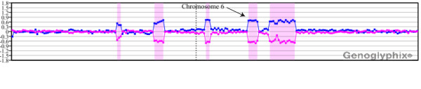
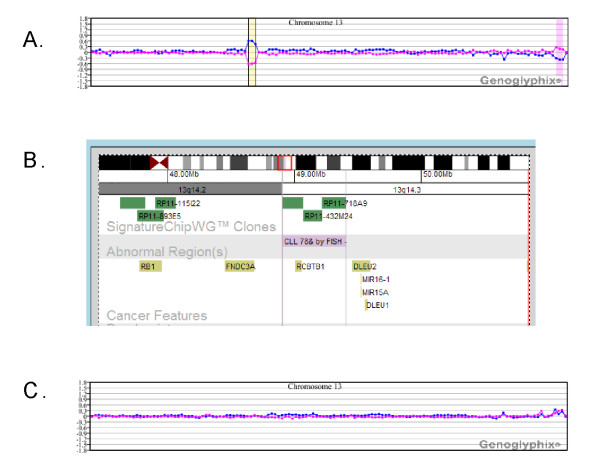
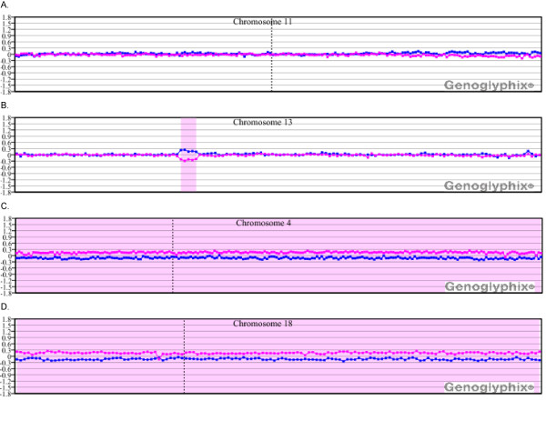
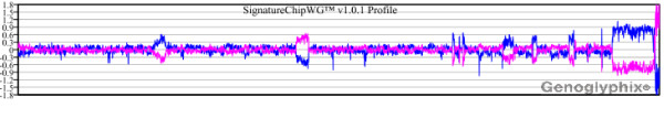
Results: We have evaluated 25 clinical cases from a tertiary cancer center that have an established diagnosis of CLL and for which there was prior cytogenetic and/or fluorescence in situ hybridization (FISH) data. We performed microarray-based comparative genomic hybridization (aCGH) using a bacterial artificial chromosome (BAC)-based microarray designed for the detection of known constitutional genetic syndromes. In 15 of the 25 cases, aCGH detected all copy number imbalances identified by prior cytogenetic and/or FISH studies. For the majority of those not detected, the aberrations were present at low levels of mosaicism. Furthermore, for 15 of the 25 cases, additional abnormalities were detected. Four of those cases had deletions that mapped to intervals implicated in inherited predisposition to CLL. For most cases, aCGH was able to detect abnormalities present in as few as 10% of cells. Although changes in ploidy are not easily discernable by aCGH, results for two cases illustrate the detection of additional copy gains and losses present within a mosaic tetraploid cell population.
Conclusions: Our results illustrate the successful evaluation of CLL using a microarray optimized for the interrogation of inherited disorders and the identification of alterations with possible relevance to CLL susceptibility.
Figures


Similar articles
-
Validation of a targeted DNA microarray for the clinical evaluation of recurrent abnormalities in chronic lymphocytic leukemia.Am J Hematol. 2008 Jul;83(7):540-6. doi: 10.1002/ajh.21145. Am J Hematol. 2008. PMID: 18161787 Clinical Trial.
-
Evaluation of chronic lymphocytic leukemia by oligonucleotide-based microarray analysis uncovers novel aberrations not detected by FISH or cytogenetic analysis.Mol Cytogenet. 2011 Nov 16;4:25. doi: 10.1186/1755-8166-4-25. Mol Cytogenet. 2011. PMID: 22087757 Free PMC article.
-
Assessing karyotype precision by microarray-based comparative genomic hybridization in the myelodysplastic/myeloproliferative syndromes.Mol Cytogenet. 2010 Nov 15;3:23. doi: 10.1186/1755-8166-3-23. Mol Cytogenet. 2010. PMID: 21078186 Free PMC article.
-
Genetic features of B-cell chronic lymphocytic leukemia.Rev Clin Exp Hematol. 2000 Mar;4(1):48-72. doi: 10.1046/j.1468-0734.2000.00003.x. Rev Clin Exp Hematol. 2000. PMID: 11486330 Review.
-
Chronic lymphocytic leukemia: a clinical and molecular heterogenous disease.Cancer Genet. 2013 Mar;206(3):49-62. doi: 10.1016/j.cancergen.2013.01.003. Epub 2013 Mar 24. Cancer Genet. 2013. PMID: 23531595 Review.
Cited by
-
Genomic imbalance defines three prognostic groups for risk stratification of patients with chronic lymphocytic leukemia.Leuk Lymphoma. 2014 Apr;55(4):920-8. doi: 10.3109/10428194.2013.845882. Epub 2013 Nov 12. Leuk Lymphoma. 2014. PMID: 24047479 Free PMC article.
-
Atypical rearrangement involving 3'-IGH@ and a breakpoint at least 400 Kb upstream of an intact MYC in a CLL patient with an apparently balanced t(8;14)(q24.1;q32) and negative MYC expression.Mol Cytogenet. 2013 Feb 1;6(1):5. doi: 10.1186/1755-8166-6-5. Mol Cytogenet. 2013. PMID: 23369149 Free PMC article.
-
The chromosomal translocation t(1;6)(p35.3;p25.2), recurrent in chronic lymphocytic leukaemia, leads to RCC1::IRF4 fusion.Br J Haematol. 2024 Dec;205(6):2321-2326. doi: 10.1111/bjh.19790. Epub 2024 Oct 15. Br J Haematol. 2024. PMID: 39406248 Free PMC article.
-
Detection of chromothripsis-like patterns with a custom array platform for chronic lymphocytic leukemia.Genes Chromosomes Cancer. 2015 Nov;54(11):668-80. doi: 10.1002/gcc.22277. Epub 2015 Aug 25. Genes Chromosomes Cancer. 2015. PMID: 26305789 Free PMC article.
-
A Japanese case of chronic lymphocytic leukemia with t (1;6).Exp Hematol Oncol. 2012 Sep 12;1(1):28. doi: 10.1186/2162-3619-1-28. Exp Hematol Oncol. 2012. PMID: 23210523 Free PMC article.
References
-
- Ries LA, Eisner MP, Kosary CL, Hankey BF, Miller BA, Clegg L, Mariotto A, Fay MP, Feuer EJ, Edwards BK. Cancer Statistics Review, 1975-2000, National Cancer Institute. Bethesda, MD; 2003.
-
- Tefferi A, O'Brien S. Chronic lymphocytic leukemia in 2007: prognostic factors and therapeutic approaches. Commun Oncol. 2007;4(suppl 5):11–16.
-
- Miller DT, Adam MP, Aradhya S, Biesecker LG, Brothman AR, Carter NP, Church DM, Crolla JA, Eichler EE, Epstein CJ, Faucett WA, Feuk L, Friedman JM, Hamosh A, Jackson L, Kaminsky EB, Kok K, Krantz ID, Kuhn RM, Lee C, Ostell JM, Rosenberg C, Scherer SW, Spinner NB, Stavropoulos DJ, Tepperberg JH, Thorland EC, Vermeesch JR, Waggoner DJ, Watson MS. et al.Consensus statement: chromosomal microarray is a first-tier clinical diagnostic test for individuals with developmental disabilities or congenital anomalies. Am J Hum Genet. 2010;86:749–764. doi: 10.1016/j.ajhg.2010.04.006. - DOI - PMC - PubMed
LinkOut - more resources
Full Text Sources
