. 2011 Apr 1;81(7):881-90.
doi: 10.1016/j.bcp.2011.01.017. Epub 2011 Feb 1.

Aurora inhibitor MLN8237 in combination with docetaxel enhances apoptosis and anti-tumor activity in mantle cell lymphoma

Affiliations

Aurora inhibitor MLN8237 in combination with docetaxel enhances apoptosis and anti-tumor activity in mantle cell lymphoma

Wenqing Qi et al. Biochem Pharmacol. .

Abstract

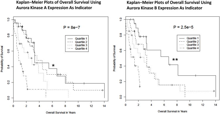
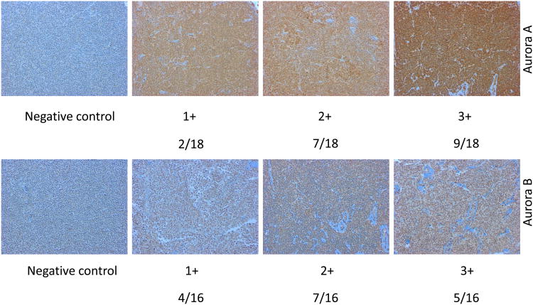
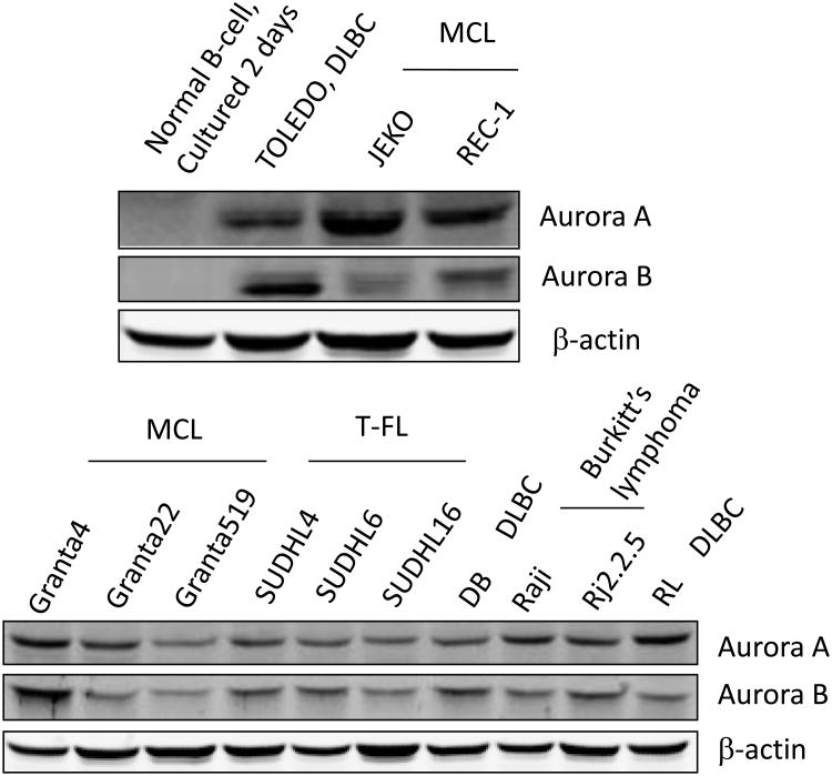
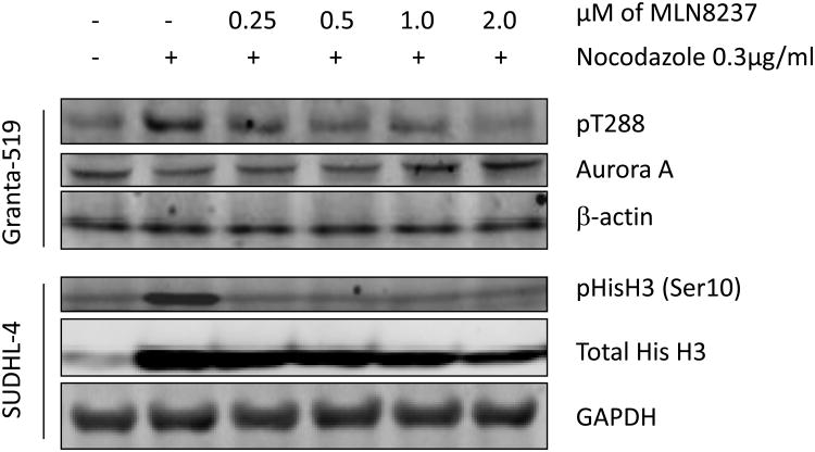
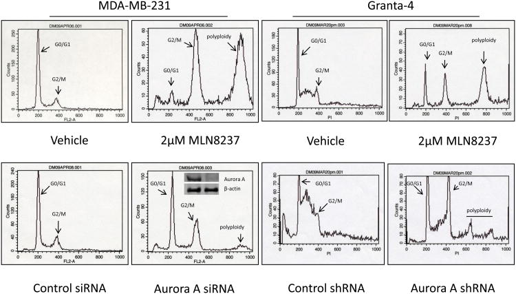
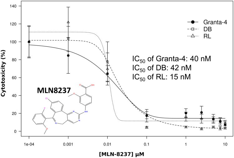
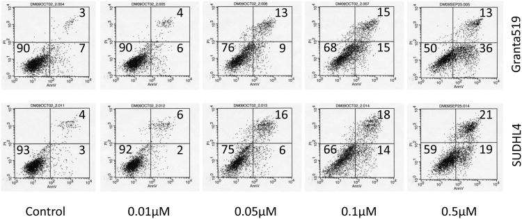
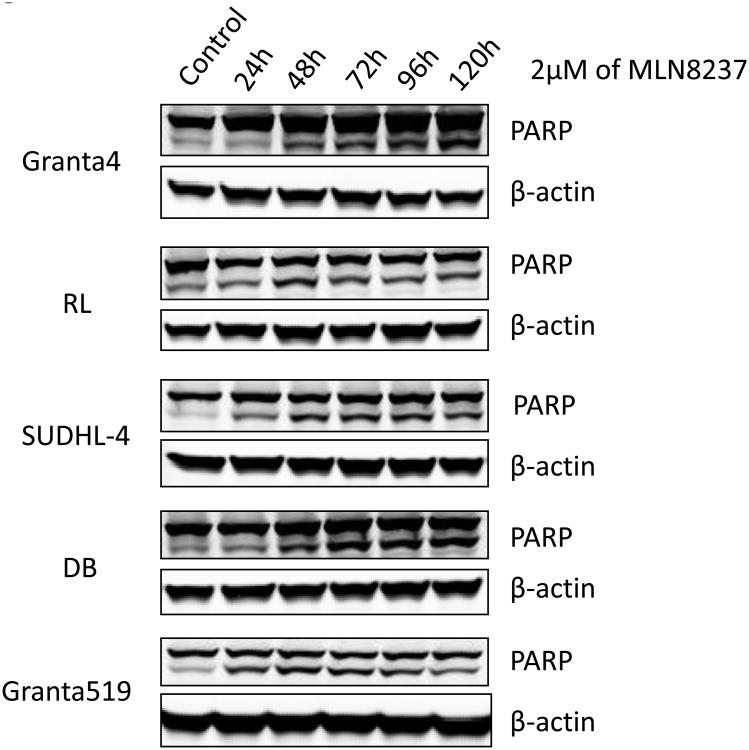
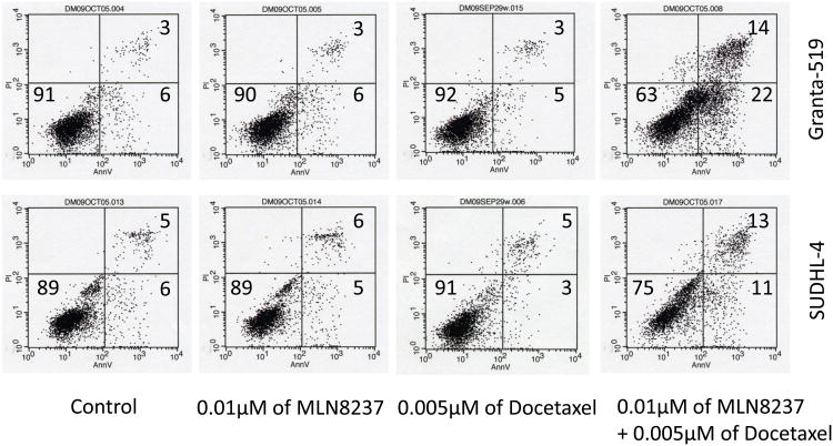
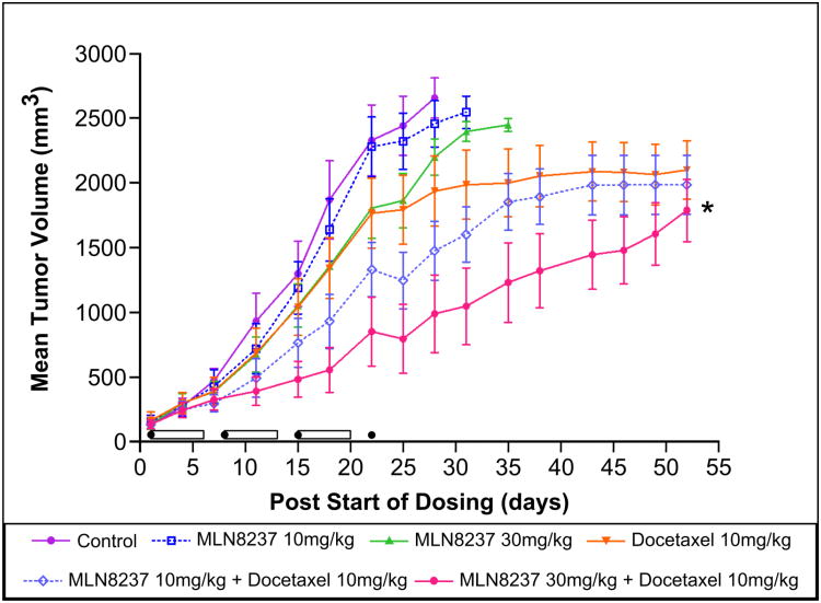
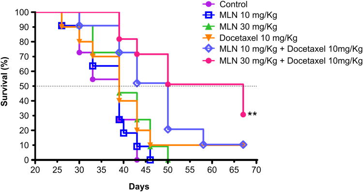
Auroras (A and B) are oncogenic serine/threonine kinases that play key roles in the mitotic phase of the eukaryotic cell cycle. Analysis of the leukemia lymphoma molecular profiling project (LLMPP) database indicates Aurora over-expression correlates with poor prognosis. A tissue microarray (TMA) composed of 20 paired mantle cell lymphoma (MCL) patients demonstrated >75% of patients had high levels Aurora expression. Aurora A and B were also found elevated in 13 aggressive B-NHL cell lines. MLN8237, an Aurora inhibitor induced G2/M arrest with polyploidy and abrogated Aurora A and histone-H3 phosphorylation. MLN8237 inhibited aggressive B-NHL cell proliferation at an IC(50) of 10-50 nM and induced apoptosis in a dose- and time-dependent manner. Low dose combinations of MLN8237+docetaxel enhanced apoptosis by ~3-4-fold in cell culture compared to single agents respectively. A mouse xenograft model of MCL demonstrated that MLN8237 (10 or 30 mg/kg) or docetaxel (10mg/kg) alone had modest anti-tumor activity. However, MLN8237 plus docetaxel demonstrated a statistically significant tumor growth inhibition and enhanced survival compared to single agent therapy. Together, our results suggest that MLN8237 plus docetaxel may represent a novel therapeutic strategy that could be evaluated in early phase trials in relapsed/refractory aggressive B-cell NHL.


Figures

Figure 1
Figure 1. Aurora A and B are over-expressed poor prognosis markers in MCL
(A) Kaplan-Meier survival curves for patients with Mantle Cell Lymphoma based on the gene expression profile obtained from the LLMPP for Aurora kinase over-expressers (quartile 4) versus the lowest expressers (quartile 1) showing high expressers have a worse survival (months) for Aurora A (* p=8.0e-7) and B (** p=2.5e-5). (B) A representative TMA IHC staining demonstrating negative control, 1+, 2+ and 3+ staining for Aurora A and B (magnification 200× for all panels) by IHC. Positive Aurora (A or B) staining/total population evaluated is shown beneath each IHC panel. (C) Western blotting analysis demonstrated that Aurora A and B are over-expressed in a panel of aggressive B-cell NHL cell lines compared to the normal B-cells.
Figure 2
Figure 2. MLN8237 inhibits Aurora A and B kinase activity and promotes polyploidy
(A) Interactive docking studies of MLN8237 with the crystal structures of the active forms of Aurora A (color by atom type) and B (green) show binding mode and score that predicts for MLN8237 to interact with both enzyme active sites with Aurora A > B. (B) Granta-519 and SUDHL-4 cells were synchronized with nocodazole without or with various concentrations of MLN8237 for 16 h. pThr288, Aurora A, pHisH3 (Ser10) and HisH3 were analyzed by Western blotting. GAPDH and β-actin were used as loading controls. (C) MLN8237 treatment results in cellular phenotypes consistent with Aurora A deficiency. Top, DNA profiles of MDA-MB-231 and Granta-4 cells treated with vehicle or MLN8237 at 2 μM for 72 h and evaluated by flow cytometry. Bottom, MDA-MB-231 and Granta-4 cells were transfected or infected by Aurora A siRNA or shRNA lentiviral particles, respectively and DNA contents were analyzed by flow cytometry at 96 h. Inset: The levels of Aurora A expression was determined for control and Aurora A knock-down cells by Western blotting analysis.
Figure 3
Figure 3. MLN8237 inhibits aggressive B-NHL cell viability and induces apoptosis
(A) Granta-4, DB and RL cells were exposed to varying concentrations of MLN8237 for 7 days. Cell viability was assessed by MTS analysis. Points are the means of triplicate determinations ± SD. Inset: IC50 values of MNL8237 in Granta-4, DB and RL cells. (B) Granta-519 and SUDHL-4 cells were treated with vehicle (control) or MLN8237 at various concentrations for 72h. Apoptosis was detected by flow cytometric analysis based on propidium iodide (Y-axis) and annexin V staining (X-axis). Percentages of survival (the lower left quadrant) and apoptotic cells (the lower and upper right quadrants) are indicated. (C) Granta-4, Granta-519, SUDHL-4, RL and DB cells were treated with MLN8237 at 2 μM for 24 h, 48 h, 72 h, 96 h and 120 h. Apoptosis was evaluated with PARP cleavage utilizing Western blotting analysis.
Figure 4
Figure 4. MLN8237 plus docetaxel abrogates mitotic delay and enhances apoptosis
Granta-519 and SUDHL-4 cells were treated with a low dose of MLN8237 (10 nM) or docetaxel (5 nM) alone or in combination for 72h. Apoptosis was analyzed by flow cytometry after annexin V and PI staining which showed a ∼3-4-fold higher apoptotic fraction with combination therapy compared to single agent therapy.
Figure 5
Figure 5. MLN8237 plus docetaxol inhibits tumor growth in a MCL xenograft mouse model and increases survival
(A). A MCL Granta-519 xenograft mouse model (n=12 per cohort) shows significant TGI in mice treated with MLN8237 plus docetaxel in a dose-dependent manner in comparison to control (* p < 0.001), MLN8237 or docetaxel alone. The cylinders represent duration of therapy with MLN8237 while the black sphere represents treatment with docetaxel. (B). A MCL Granta-519 xenograft mouse model showed significant overall survival difference with MLN8237 plus docetaxel in comparison to control (** p=0.0003), MLN8237 or docetaxel alone.

Similar articles

Cited by

References

    1. Marumoto T, Zhang D, Saya H. Aurora-A - a guardian of poles. Nat Rev Cancer. 2005;5:42–50. - PubMed
    1. Nigg EA. Mitotic kinases as regulators of cell division and its checkpoints. Nat Rev Mol Cell Biol. 2001;2:21–32. - PubMed
    1. Crosio C, Fimia GM, Loury R, Kimura M, Okano Y, Zhou H, et al. Mitotic phosphorylation of histone H3: spatio-temporal regulation by mammalian Aurora kinases. Mol Cell Biol. 2002;22:874–85. - PMC - PubMed
    1. Giet R, Prigent C. Aurora/Ipl1p-related kinases, a new oncogenic family of mitotic serine-threonine kinases. J Cell Sci. 1999;112(Pt 21):3591–601. - PubMed
    1. Kufer TA, Nigg EA, Sillje HH. Regulation of Aurora-A kinase on the mitotic spindle. Chromosoma. 2003;112:159–63. - PubMed

Publication types

MeSH terms