Review
. 2011 Feb 8;57(6):641-52.
doi: 10.1016/j.jacc.2010.11.010.

Clinical, molecular, and genomic changes in response to a left ventricular assist device

Affiliations
Review

Clinical, molecular, and genomic changes in response to a left ventricular assist device

Jennifer L Hall et al. J Am Coll Cardiol. .

Abstract

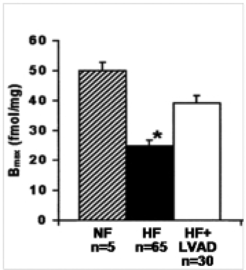
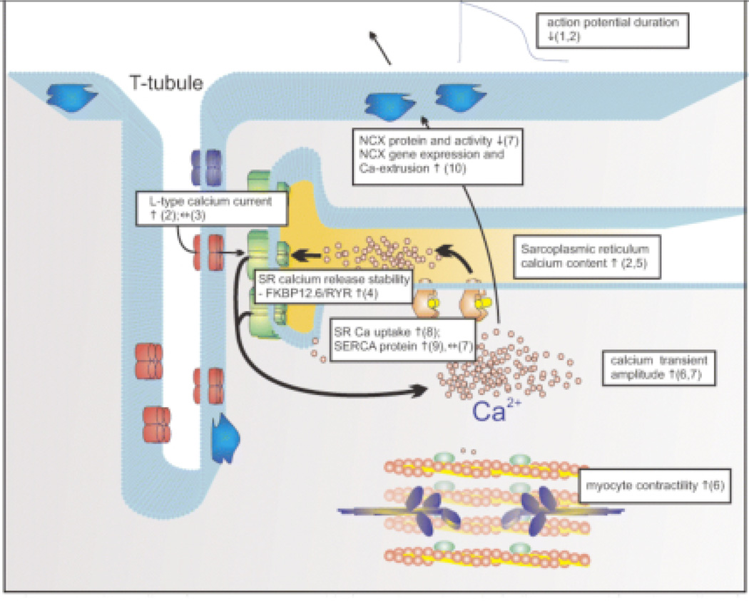
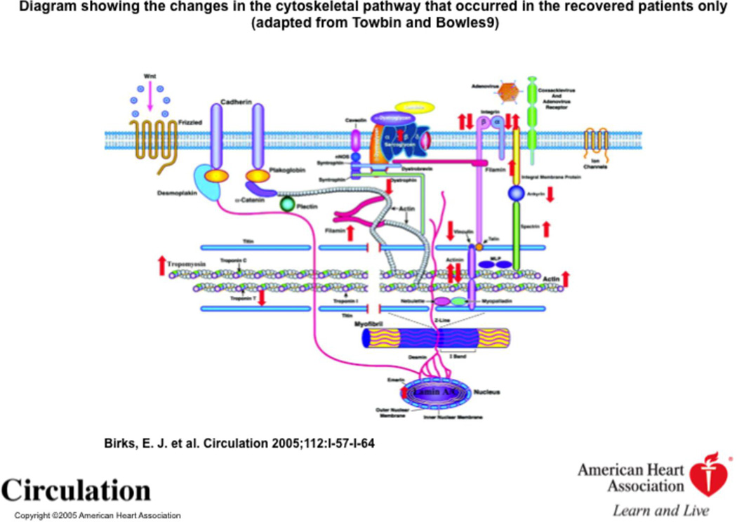
The use of left ventricular assist devices in treating patients with end-stage heart failure has increased significantly in recent years, both as a bridge to transplantation and as destination therapy in those who are ineligible for cardiac transplantation. This increase is based largely on the results of several recently completed clinical trials with the new second-generation continuous-flow devices that showed significant improvements in survival, functional capacity, and quality of life. Additional information on the use of the first- and second-generation left ventricular assist devices has come from a recently released report spanning the years 2006 to 2009, from the Interagency Registry for Mechanically Assisted Circulatory Support, a National Heart, Lung, and Blood Institute-sponsored collaboration between the U.S. Food and Drug Administration, the Centers for Medicare and Medicaid Services, and the scientific community. The authors review the latest clinical trials and data from the registry, with tight integration of the landmark molecular, cellular, and genomic research that accompanies the reverse remodeling of the human heart in response to a left ventricular assist device and functional recovery that has been reported in a subset of these patients.


Figures

Figure 1
Figure 1
Response of muscles to 1 µmol/L isoproterenol. Muscles from nonfailing hearts (NF), failing hearts without LVAD (Failing), and failing hearts with LVAD (Failing+LVAD). A, TPT (time to peak tension); B, THR (time to half relaxation); C, +dT/dt (maximal rate of tension rise); and D, −dT/dt (maximal rate of tension fall). Data are shown as change from baseline contraction (mean±SEM). *P<0.05 vs NF. n=No. of muscles/No. of hearts. This study shows no change in time to peak tension (A) or time to half relaxation (B) between non-failing, failing hearts and failing + LVAD. A significant fall in +dT/dt (C) and −dT/dt (D) was seen in the failing hearts that was improved with the LVAD. (Adopted from Ogletree-Hughes et al, Circulation, 2001).
Figure 2
Figure 2
Density of beta-adrenergic receptors in LV and RV myocardial samples of nonfailing (ruled bars), medical (black bars), and LVAD-supported hearts (open bars). Data reported are the means ± SD of maximal binding (Bmax) values determined from Scatchard transformation of saturated binding data. *p < 0.01 vs. nonfailing and LVAD-supported hearts. (Adopted from Klotz et al, 2005).
Figure 3
Figure 3
Identification of genes in the camp-mediated signaling pathway whose expression was altered in association with recovery. Shaded symbols represent genes whose expression was significantly altered in explanted vs. implanted samples. (Hall et al, 2007).
Figure 4
Figure 4
Calcium handling following LVAD support. This figure summarizes work to date in the literature of VADs. (1) Harding JD et al., Circulation 2001; (2) Terracciano CMet al., Circulation 2004; (3) Chen X et al., Circ. Res. 2002; (4) Marx SO et al., Cell 2000; (5) Terracciano CM et al., Eur. Heart J. 2003; (6) Dipla K et al., Circulation 1998; (7) Chaudhary K et al., J. Am. Coll. Cardiol. 2004; (8) Frazier MD et al., Ann. Thor. Surg. 1999; (9) Heerdt PM et al., Circulation 2000; (10) (Terracciano et al, 2007). (Terracciano, et al, 2007).
Figure 5
Figure 5
Sarcoplasmic reticulum calcium content from myocytes isolated from left ventricular assist device cores and tissue from explanted (recovery) and transplanted hearts without recovery. (Taken from Terracciano et al, 2004).
Figure 6
Figure 6
Developed tension, expressed as percent change from baseline. (A) Stimulation delay–response curves at delay periods ranging from 2 to 120 seconds (n = number of muscles/number of hearts). (B) Data from the 40-second delay period. Right: separation of F+LVAD by duration of LVAD support. Short duration = 7 to 103 days; long duration = 127 to 334 days. (C) Relationship between duration and PRP recovery (p < 0.05) at 40-second delay in hearts with an LVAD, by heart. F, failing; NF, nonfailing; F+LVAD, LVAD-supported failing heart. *p < 0.05 vs NF. (Adopted from Ogletree et al, 2010).
Figure 7
Figure 7
EPAC2 mRNA levels are significantly reduced in recovered hearts following VAD support (n=11, p< 0.01). No significant differences were seen in non-recovered hearts. (n=5, p ns). (Hall et al, 20
Figure 8
Figure 8
Diagram showing the changes in the cytoskeletal pathway that occurred in the recovered patients only (Birks et al, 2005, adapted from Towbin and Bowles).

Comment in

References

    1. Kirklin JK, Naftel DC, Kormos RL, et al. Second INTERMACS annual report: more than 1,000 primary left ventricular assist device implants. J Heart Lung Transplant. 2010;29:1–10. - PMC - PubMed
    1. Hunt SA, Abraham WT, Chin MH, et al. 2009 focused update incorporated into the ACC/AHA 2005 Guidelines for the Diagnosis and Management of Heart Failure in Adults: a report of the American College of Cardiology Foundation/American Heart Association Task Force on Practice Guidelines: developed in collaboration with the International Society for Heart and Lung Transplantation. Circulation. 2009;119:e391–e479. - PubMed
    1. Jessup M, Brozena S. Heart failure. N Engl J Med. 2003;348:2007–2018. - PubMed
    1. Miller LW, Pagani FD, Russell SD, et al. Use of a continuous-flow device in patients awaiting heart transplantation. N Engl J Med. 2007;357:885–896. - PubMed
    1. Slaughter MS, Rogers JG, Milano CA, et al. Advanced heart failure treated with continuous-flow left ventricular assist device. N Engl J Med. 2009;361:2241–2251. - PubMed

Publication types