Review
. 2011 Feb 4;41(3):249-62.
doi: 10.1016/j.molcel.2011.01.008.

Single-molecule studies of RNA polymerase: one singular sensation, every little step it takes

Affiliations
Review

Single-molecule studies of RNA polymerase: one singular sensation, every little step it takes

Matthew H Larson et al. Mol Cell. .

Abstract

Transcription is the first of many biochemical steps that turn the genetic information found in DNA into the proteins responsible for driving cellular processes. In this review, we highlight certain advantages of single-molecule techniques in the study of prokaryotic and eukaryotic transcription, and the specific ways in which these techniques complement conventional, ensemble-based biochemistry. We focus on recent literature, highlighting examples where single-molecule methods have provided fresh insights into mechanism. We also present recent technological advances and outline future directions in the field.


Figures

Figure 1
Figure 1. Single-molecule experimental geometries and representative velocity data
(A) In the tethered particle assay, RNAP (green) is absorbed non-specifically onto a coverslip surface. As RNAP transcribes downstream (green arrow) along the DNA template (blue), the length of the tether between the bead (light blue) and RNAP shortens, reducing the Brownian excursions of the bead, which can be used to monitor the tether length. (B) In the DNA-pulling single-trap assay, the upstream end of the DNA template is tethered to a coverslip via an antibody linkage (black and purple). RNAP is attached to a polystyrene bead via a biotin-avidin linkage (black and yellow), and the bead is pulled towards an optical trap (pink) that provides a restoring force, F. (C) In the RNA-pulling double trap assay, a 3-kb DNA handle (dark blue) with a 25 nt single-stranded overhang is tethered to a larger bead via a bitoin-avidin linkage. The overhang is complementary to the 5′ end of the RNA, and is annealed to the nascent transcript. RNAP is tethered to a smaller bead via a second a biotin-avidin linkage. The beads are maintained in separate optical traps free of the coverglass surface. (D) Single-molecule transcription records: tether length is converted into position of RNAP along the template and plotted as a function of time. Some pauses are indicated (red arrows). Many records end in the immediate region of an intrinsic terminator (solid gray bar). (E) Histogram (blue bars) of the average instantaneous velocities from individual transcription records (N = 123); a Gaussian fit to these data (solid red line) supplies a mean velocity of 23 ± 11 bp/s. By comparison, the narrower distribution based on the average deviations of velocities within a given trace is shown (dotted black line). The additional variance indicates heterogeneity.
Figure 2
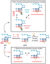
Figure 2. Model of transcriptional pausing
A Brownian ratchet model of the RNAP transcription cycle, consistent with single-molecule data for both E. coli and T7 RNAP (Abbondanzieri et al., 2005a; Thomen et al., 2008), shown relative to the nucleic acid scaffold. During elongation, RNAP shuttles back and forth between its pre- and post-translocated states until nucleotide binding rectifies this motion and an incoming NTP is incorporated into the growing transcript. Pausing represents an off-pathway event, and is depicted here as branching from the pre-translocated state. In the elemental pause model (upper panel), pausing results from fraying of the RNA 3′ end into an alternate site (green circle) located distal to the active site, and does not involve any translocation of RNAP with respect to the scaffold. Such a state can serve as a precursor to a hairpin or backtracked pause. In the reverse translocation pause model (lower panel), pausing results from a backtracking motion of RNAP along DNA through 1 or more bp. The pause duration is determined by the time taken for RNAP to diffuse back to its original starting position, where the RNA 3′ end realigns with the active site.
Figure 3
Figure 3. Pol II factorless initiation and the single-molecule assay geometry
To form the RNA:DNA hybrid, a short synthetic RNA transcript (red) is first hybridized to a DNA oligo (light blue) that serves as the template strand. Then, a biotin-labeled Pol II molecule (green) is bound to this RNA:DNA hybrid, after which a complementary non-template strand (dark blue) is added to form a stalled, but functional, elongation complex. Long, dsDNA molecules (purple) are subsequently ligated to both the upstream and downstream ends of the pre-fabricated scaffold. The upstream dsDNA handle allows for ∼1 μm of separation between the two beads, while the downstream dsDNA serves as the template for transcription, and includes (in this case) a nucleosome positioning sequence (NPS). The entire complex is suspended between two polystyrene beads, and tension is exerted in the tether in a manner that assists Pol II during translocation (green arrow).
Figure 4
Figure 4. The effect of the U-rich tract on transcription termination
(A) Plot of termination efficiency (TE) as a function of force exerted on RNAP. For the λtR2 tetraloop terminator (red squares), which has a single interruption in its U-tract, TE is flat over the accessible force range. For the t500 terminator (green circles), which has two interruptions in its U-tract, TE increases with force. (B) Fusion of the λtR2 hairpin with the t500 U-tract creates a chimeric terminator that is sensitive to force (blue circles). (C) A model for the termination pathway. When RNAP reaches an intrinsic terminator sequence with a perfect or near-perfect U-tract (upper pathway), hairpin closure pulls the RNA away from the RNAP enzyme, causing the RNA to shear against template DNA in the hybrid without causing forward translocation of RNAP. In the case of an interrupted U-tract (lower pathway), RNAP must first forward translocate by ∼1.4 bp, effectively shortening and de-stabilizing the RNA:DNA hybrid prior to leading to the release of the transcript.
Figure 5
Figure 5. Single-molecule, FRET-based nanopositioning assay
(A) RNAP (green) is labeled with a donor dye (blue star). The donor dye is excited by an external light source (purple arrows) and transfers its excitation to the acceptor dye (orange star) via FRET (blue arrow). The intensity of the FRET signal is proportional to the inverse sixth power of the distance between the dyes. (B) By placing multiple donor dyes on both the RNAP and the DNA template, it becomes possible to triangulate the location of the mobile element carrying the acceptor dye by means of multiple FRET measurements of intramolecular distances (light blue circles).

References

    1. Abbondanzieri EA, Greenleaf WJ, Shaevitz JW, Landick R, Block SM. Direct observation of base-pair stepping by RNA polymerase. Nature. 2005a;438:460–465. - PMC - PubMed
    1. Abbondanzieri EA, Shaevitz JW, Block SM. Picocalorimetry of transcription by RNA polymerase. Biophys J. 2005b;89:L61–63. - PMC - PubMed
    1. Adelman K, La Porta A, Santangelo TJ, Lis JT, Roberts JW, Wang MD. Single molecule analysis of RNA polymerase elongation reveals uniform kinetic behavior. Proc Natl Acad Sci U S A. 2002;99:13538–13543. - PMC - PubMed
    1. Allison LA, Moyle M, Shales M, Ingles CJ. Extensive homology among the largest subunits of eukaryotic and prokaryotic RNA polymerases. Cell. 1985;42:599–610. - PubMed
    1. Andrecka J, Lewis R, Bruckner F, Lehmann E, Cramer P, Michaelis J. Single-molecule tracking of mRNA exiting from RNA polymerase II. Proc Natl Acad Sci U S A. 2008;105:135–140. - PMC - PubMed

LinkOut - more resources