. 2011 Feb 4;41(3):286-97.
doi: 10.1016/j.molcel.2010.12.027.

The base-pairing RNA spot 42 participates in a multioutput feedforward loop to help enact catabolite repression in Escherichia coli

Affiliations

The base-pairing RNA spot 42 participates in a multioutput feedforward loop to help enact catabolite repression in Escherichia coli

Chase L Beisel et al. Mol Cell. .

Abstract

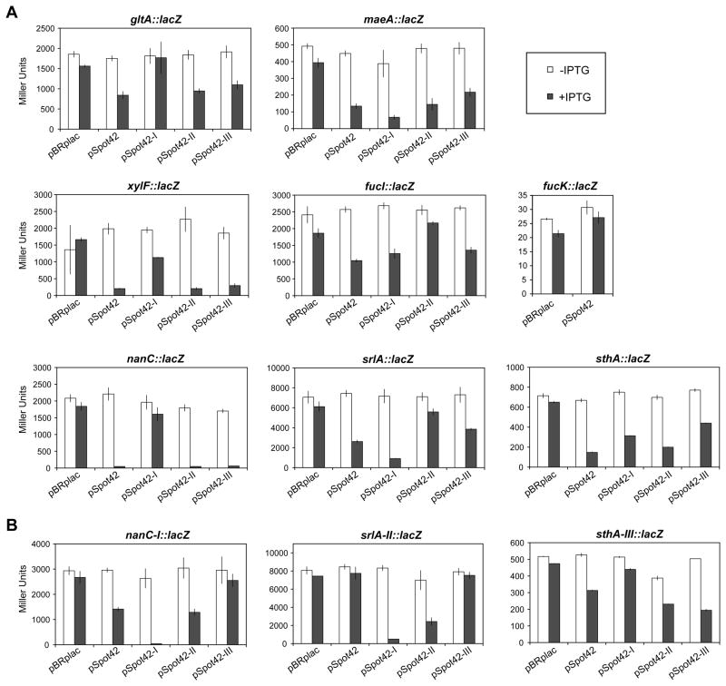
Bacteria selectively consume some carbon sources over others through a regulatory mechanism termed catabolite repression. Here, we show that the base-pairing RNA Spot 42 plays a broad role in catabolite repression in Escherichia coli by directly repressing genes involved in central and secondary metabolism, redox balancing, and the consumption of diverse nonpreferred carbon sources. Many of the genes repressed by Spot 42 are transcriptionally activated by the global regulator CRP. Since CRP represses Spot 42, these regulators participate in a specific regulatory circuit called a multioutput feedforward loop. We found that this loop can reduce leaky expression of target genes in the presence of glucose and can maintain repression of target genes under changing nutrient conditions. Our results suggest that base-pairing RNAs in feedforward loops can help shape the steady-state levels and dynamics of gene expression.


Figures

Figure 1
Figure 1
Repression of gltA, maeA, sthA, and srlA following Spot 42 overexpression. The Spot 42 gene spf was cloned downstream of an IPTG-inducible promoter in the plasmid pBRplac, yielding pSpot42. NM525 Δspf::kanR (GS0433) cells transformed with pBRplac or pSpot42 were grown in (A) LB or (B) LB containing 0.2% D-sorbitol to an ABS600 of ~0.3 and treated with 1 mM IPTG. After different periods of time, total RNA was isolated and subjected to northern blot analysis with probes for (A) gltA, maeA, sthA, and (B) srlA. 5S RNA served as a loading control. Results are representative of two independent experiments.
Figure 2
Figure 2. Mutational analysis of base-pairing interactions between Spot 42 and target mRNAs
(A) Results for β-galactosidase assays of PM1205 Δspf::kanR (GS0434) cells containing lacZ translational fusions. (B) Results for β-galactosidase assays for lacZ translational fusions with compensatory mutations in the predicted location of base pairing with Spot 42. Derivatives of PM1205 Δspf::kanR transformed with the indicated plasmid were grown in LB to an ABS of ~0.1 and treated with 0.2% arabinose or 0.2% arabinose and 1 mM IPTG for 1 h before being subjected to β-galactosidase assays. The mutations in pSpot42 variant I – III correspond to the indicated mutations in Figure 3A. The compensatory mutations in the lacZ translational fusions correspond to the indicated mutations in Figure 3C. The reported averages and standard deviations are from measurements of cultures from three separate colonies. See Figure S1 for predicted base-pairing interactions between Spot 42 variant I and the srlA::lacZ and maeA::lacZ fusions.
Figure 3
Figure 3. Predicted base-pairing interactions between the single-stranded regions of Spot 42 and target mRNAs
(A) Secondary structure of Spot 42 reported previously (Møller et al., 2002b). The three single-stranded regions are highlighted in gray. Three consecutive nucleotides (white) in each single-stranded region were mutated to disrupt predicted base-pairing interactions with target mRNAs (I – III). (B) Northern blot analysis of NM525 Δspf::kanR (GS0433) cells transformed with pBRplac, pSpot42, or pSpot42 variants I – III. Cells were grown in LB to an ABS600 of ~0.3, treated with 1 mM IPTG, and incubated for 0 min or 30 min. An equimolar concentration of probes spf.north1 and spf.north2 were used to detect all Spot 42 variants on the same membrane. 5S RNA served as a loading control. (C) Genes identified by microarray analysis and base-pairing interactions with Spot 42 predicted by the folding algorithm NUPACK. NUPACK did not predict any significant base-pairing interactions between Spot 42 and maeA. Mutations introduced into pSpot42 and the lacZ translational fusions are designated. The bar below each target gene indicated as a darker arrow designates the predicted location of base pairing with Spot 42. The number above each promoter specifies the number of nucleotides between a transcriptional start site and the start codon of the first gene in the operon.
Figure 4
Figure 4. Limited growth on non-preferred carbon sources following Spot 42 overexpression
NM525 Δspf::kanR (GS0433) cells transformed with pBRplac or pSpot42 were grown overnight in LB or in casamino acid-enriched M9 containing the indicated carbon source and diluted to an ABS600 of 0.01 into the same type of media with or without 1 mM IPTG. ABS600 was measured at different times during cell growth. Applied concentration of specific carbon sources: 0.4% glycerol, 0.8% sodium succinate hexahydrate, 0.2% L-fucose, 2 mM N-acetylneuraminic acid (Neu5Ac), 0.2% D-sorbitol, 0.2% D-xylose, and 60 mM L-lactic acid. Results are representative of two independent experiments. See Figure S2 for growth data for extended time periods, for media lacking casamino acids, and for strains containing a deletion of Spot 42 target genes.
Figure 5
Figure 5. Steady-state behavior of the CRP-Spot 42 feedforward loop
(A) CRP and Spot 42 participate in a multi-output coherent feedforward loop. CRP positively regulates target expression directly at the transcriptional level and indirectly by repressing Spot 42. Mutations were introduced into the wild type SPA fusion strain (wt) that reduce the loop to direct regulation by CRP (Δspf) or eliminate regulation by both CRP and Spot 42 (Δspf Δcrp). (B) Results from quantitative western blot analysis for srlA-SPA strains. (C) Results from quantitative western blot analysis for fucI-SPA strains. For both B and C, strains were grown in LB or LB containing 0.2% glucose. Northern blot analysis was used to detect the levels of Spot 42. GroEL and 5S RNA served as loading controls. Results are representative of two independent experiments. See Figure S3 for dilution series, for data for cells grown in casamino acid-enriched M9 containing glycerol or casamino acid-enriched M9 containing glycerol and glucose, for β-galactosidase assay results for srlA-lacZ and fucI-lacZ fusion strains, and steady-state data for strains containing a destabilized version of SrlA-SPA.
Figure 6
Figure 6. Regulatory dynamics of the CRP-Spot 42 feedforward loop
(A) Time course for SrlA-SPA levels following deactivation of CRP. srlA-SPA strains containing the full loop (wt) or the loop reduced to direct regulation by CRP (Δspf) were grown in casamino acid-enriched M9 containing 0.4% glycerol in multiple tubes and 0.2% glucose was added to each tube at the indicated time prior to harvesting the cultures. Normalized protein levels calculated from quantitative western blot analysis were rescaled to span 0 to 100. (B) Time course for SrlA-SPA levels following activation of CRP. wt and Δspf strains were grown in casamino acid-enriched M9 containing 0.4% glycerol and 0.2% glucose in multiple tubes and 10 mM cAMP was added to each tube at the indicated time prior to harvesting the cultures. (C) Time course for FucI-SPA levels following deactivation of CRP. fucI-SPA strains containing the full loop (wt) or the loop reduced to direct regulation by CRP (Δspf) were grown as described in A. (D) Time course for FucI-SPA levels following activation of CRP. wt and Δspf strains were grown as described in B. Results are representative of three independent experiments. See Figure S4 for dilution series, for an independent set of time course data, for growth rates of the srlA-SPA and fucI-SPA strains, and for time course data for strains expressing a destabilized version of SrlA-SPA.

Comment in

References

    1. Aiba H, Adhya S, de Crombrugghe B. Evidence for two functional gal promoters in intact Escherichia coli cells. J Biol Chem. 1981;256:11905–11910. - PubMed
    1. Alon U. Network motifs: theory and experimental approaches. Nat Rev Genet. 2007;8:450–461. - PubMed
    1. Ansong C, Yoon H, Porwollik S, Mottaz-Brewer H, Petritis BO, Jaitly N, Adkins JN, McClelland M, Heffron F, Smith RD. Global systems-level analysis of Hfq and SmpB deletion mutants in Salmonella: implications for virulence and global protein translation. PLoS One. 2009;4:e4809. - PMC - PubMed
    1. Beisel CL, Storz G. Base pairing small RNAs and their roles in global regulatory networks. FEMS Microbiol Rev. 2010;34:866–882. - PMC - PubMed
    1. Boysen A, Møller-Jensen J, Kallipolitis B, Valentin-Hansen P, Overgaard M. Translational regulation of gene expression by an anaerobically induced small non-coding RNA in Escherichia coli. J Biol Chem. 2010;285:10690–10702. - PMC - PubMed

Publication types