. 2011 Feb 4;41(3):311-20.
doi: 10.1016/j.molcel.2011.01.013.

Structural basis for dimerization and activity of human PAPD1, a noncanonical poly(A) polymerase

Affiliations

Structural basis for dimerization and activity of human PAPD1, a noncanonical poly(A) polymerase

Yun Bai et al. Mol Cell. .

Abstract

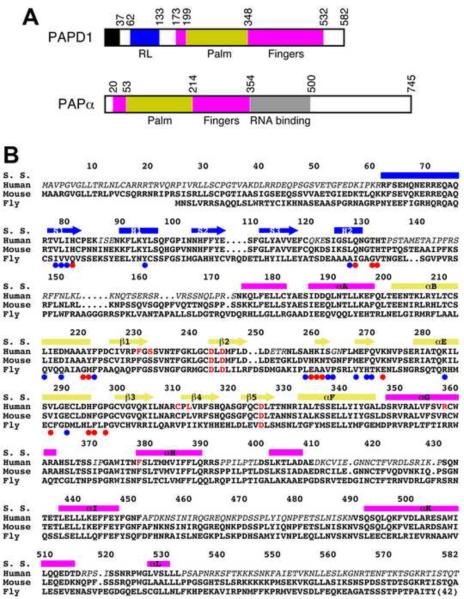
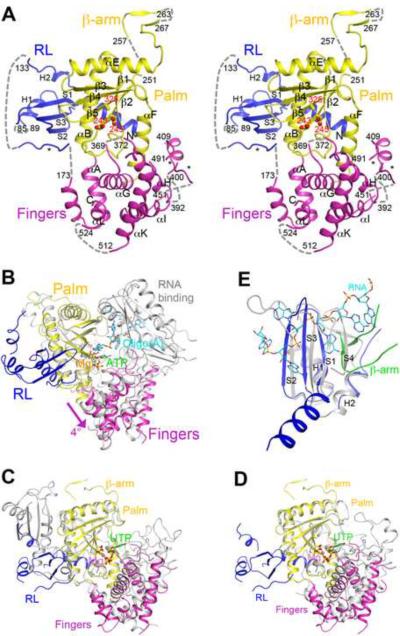
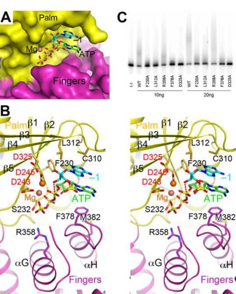
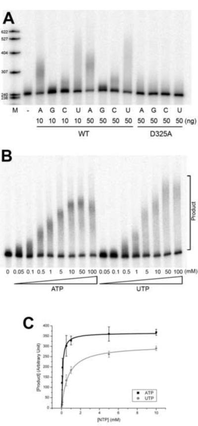
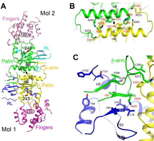
Poly(A) polymerases (PAPs) are found in most living organisms and have important roles in RNA function and metabolism. Here, we report the crystal structure of human PAPD1, a noncanonical PAP that can polyadenylate RNAs in the mitochondria (also known as mtPAP) and oligouridylate histone mRNAs (TUTase1). The overall structure of the palm and fingers domains is similar to that in the canonical PAPs. The active site is located at the interface between the two domains, with a large pocket that can accommodate the substrates. The structure reveals the presence of a previously unrecognized domain in the N-terminal region of PAPD1, with a backbone fold that is similar to that of RNP-type RNA binding domains. This domain (named the RL domain), together with a β-arm insertion in the palm domain, contributes to dimerization of PAPD1. Surprisingly, our mutagenesis and biochemical studies show that dimerization is required for the catalytic activity of PAPD1.


Figures

Figure 1
Figure 1
Sequence conservation of PAPD1. (A). Schematic drawing of the domain organization of human PAPD1 and human PAPα. The various domains are labeled. The mitochondrial targeting sequence of PAPD1 is shown in black. (B). Sequence alignment of human, murine and Drosophila melanogaster (Fly, CG11418) PAPD1. The secondary structure elements (S. S.) are labeled and colored according to the domains. Residues shown in red are in the active site. The colored dots highlight those residues with >50 Å2 (red) or >20 Å2 (blue) buried surface area in the dimer interface. Residues missing in the current structure of human PAPD1 are shown in italic. The sequence conservation between human and Drosophila PAPD1 for residues in the RL domain (25% identity) is lower compared to that for the palm and fingers domains (39% identity).
Figure 2
Figure 2
Structure of human PAPD1 monomer. (A). Stereo drawing of the structure of human PAPD1 monomer. The two catalytic Asp residues and the D325A mutation in the active site are shown in stick models and labeled in red. Three segments modeled as poly-alanines are indicated with the stars. The missing segments of the structure are indicated with the dashed lines (gray). (B). Overlay of the structure of human PAPD1 (in color) with that of yeast PAP (in gray) in complex with Mg2+ (pink sphere), ATP (green) and oligo(A) (cyan) (Balbo and Bohm, 2007). The fingers domain of PAPD1 is in a slightly more open conformation compared to that in PAP, indicated with the arrow (magenta). (C). Overlay of the structure of human PAPD1 (in color) and T. brucei TUTase2 (in gray) (Deng et al., 2005). The inserted domain in the palm domain of TUTase2 is in a different location compared to the RL domain of PAPD1. (D). Overlay of the structure of human PAPD1 (in color) and T. brucei TUTase4 (in gray) (Stagno et al., 2007a; Stagno et al., 2007b). For stereo versions of panels C and D, please see Supplementary Fig. 3. (E). Overlay of the RL domain of human PAPD1 (in blue) with the first RRM of PABP (in gray) in complex with oligo(A) (cyan) (Deo et al., 1999). The β-arm of the second monomer of PAPD1 is shown in green, and it clashes with the fourth strand of the RRM. All the structure figures were produced with PyMOL (www.pymol.org). See also Figs. S1–3.
Figure 3
Figure 3
Model of the active site of human PAPD1 with substrates. (A). Molecular surface of the active site region of PAPD1. The bound positions of Mg2+ (pink sphere), ATP (green) and the last nucleotide of the RNA substrate (cyan, labeled −1) are modeled from the structure of yeast PAP (Balbo and Bohm, 2007). (B). Stereo drawing of the active site model of human PAPD1. Side chains with potentially important roles in catalysis and/or substrate binding are shown as stick models and labeled. The side chain of Asp325 and the two Mg2+ ions are modeled. The inline nucleophilic attack of the 3' hydroxyl group of the last nucleotide of the RNA substrate (the −1 nucleotide) on the α-phosphate of ATP, modeled based on the structure of yeast PAP (Balbo and Bohm, 2007), is indicated by the dashed line in red. (C). Poly(A) polymerase assays for active site mutants of PAPD1. 10 or 20 ng of each enzyme was used in the assay, and the D325A mutant was included as a control.
Figure 4
Figure 4
PAPD1 has activity towards all four nucleotides in vitro. (A). Catalytic activity of PAPD1 towards the four nucleotides, ATP, GTP, CTP and UTP. 10 and 50 ng of the wild-type (WT) enzyme were used, and 50 ng of the D325A mutant were also assayed as a control. (B). Concentration titration of the activity of PAPD1 towards ATP and UTP. The concentrations used for the nucleotides are indicated. (C). The poly(A) and poly(U) polymerase reaction of PAPD1 follows Michaelis-Menten kinetics. The error bars represent ± 1SD.
Figure 5
Figure 5
The dimer of human PAPD1. (A). Schematic drawing of the human PAPD1 dimer. The domains in the two monomers are given different colors and labeled, and Mol 1 is the one shown in Fig. 2A. The catalytic Asp residues in the active sites are shown in stick models. The two-fold axis of the dimer is indicated with the oval in black. (B). Detailed interactions between helices αE and αB in one contact area of the dimer interface. Residues labeled in red have been selected for mutagenesis experiments. (C). Detailed interactions between the RL domain of one monomer and the β-arm of the other monomer in the other contact area of the dimer interface. See also Fig. S4.
Figure 6
Figure 6
Dimerization is required for the catalytic activity of PAPD1. (A). Gel-filtration data showing that a sextuple mutation in the dimer interface, H259A/K260A/I261A/H294A/F295A/P297A, produces a stable monomer in solution. The profile for wild-type PAPD1 is shown in black, and that for the mutant in gray. The void volume on this column is at 7.8 ml, aldolase (158 kD) at 12.1 ml, and albumin (67 kD) at 13.4 ml. (B). Poly(A) polymerase assay for wild-type PAPD1 (residues 44–538), the D325A active-site mutant, and the sextuple mutant H259A/K260A/I261A/H294A/F295A/P297A (labeled monomeric mutant).

References

    1. Adams PD, Grosse-Kunstleve RW, Hung L-W, Ioerger TR, McCoy AJ, Moriarty NW, Read RJ, Sacchettini JC, Sauter NK, Terwilliger TC. PHENIX: building a new software for automated crystallographic structure determination. Acta Cryst. 2002;D58:1948–1954. - PubMed
    1. Aravind L, Koonin EV. DNA polymerase b-like nucleotidyltransferase superfamily: identification of three new families, classification and evolutionary history. Nucl Acid Res. 1999;27:1609–1618. - PMC - PubMed
    1. Balbo PB, Bohm A. Mechanism of poly(A) polymerase: structure of the enzyme-MgATP-RNA ternary complex and kinetic analysis. Structure. 2007;15:1117–1131. - PMC - PubMed
    1. Balbo PB, Meinke G, Bohm A. Kinetic studies of yeast polyA polymerase indicate an induced fit mechanism for nucleotide specificity. Biochem. 2005;44:7777–7786. - PubMed
    1. Balbo PB, Toth J, Bohm A. X-ray crystallographic and steady state fluorescence characterization of the protein dynamics of yeast polyadenylate polymerase. J Mol Biol. 2007;366:1401–1415. - PMC - PubMed

Publication types

Substances