Review
. 2011 Mar 15;411(2):362-73.
doi: 10.1016/j.virol.2010.12.045. Epub 2011 Feb 2.

Complement and viral pathogenesis

Affiliations
Review

Complement and viral pathogenesis

Kristina A Stoermer et al. Virology. .

Abstract

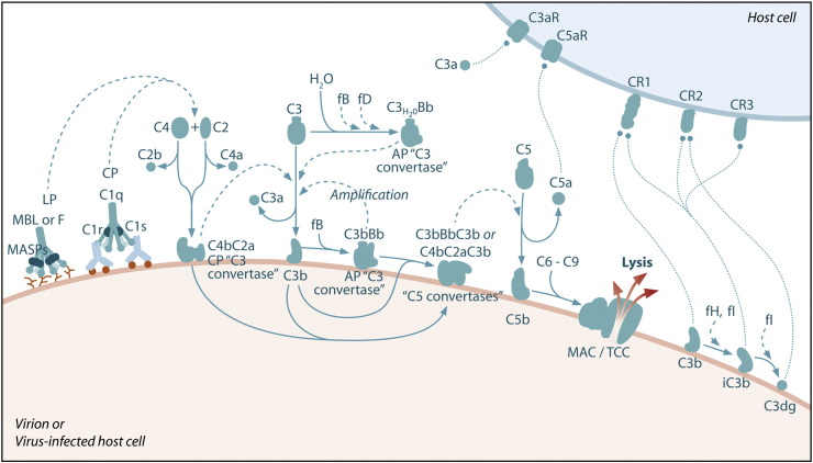
The complement system functions as an immune surveillance system that rapidly responds to infection. Activation of the complement system by specific recognition pathways triggers a protease cascade, generating cleavage products that function to eliminate pathogens, regulate inflammatory responses, and shape adaptive immune responses. However, when dysregulated, these powerful functions can become destructive and the complement system has been implicated as a pathogenic effector in numerous diseases, including infectious diseases. This review highlights recent discoveries that have identified critical roles for the complement system in the pathogenesis of viral infection.


Figures

Fig. 1
Fig. 1
Schematic of the complement system. Complement is activated by three major pathways. The classical pathway is primarily activated when C1q interacts with IgM and certain IgG isotypes bound to antigen. C1q-associated C1s cleaves C4 and C2 to form the classical pathway (CP) C3 convertase (C4bC2a). The lectin pathway (LP) is initiated by carbohydrate pattern recognition receptors such as mannose-binding lectin (MBL) and the ficolins (F) which are in a complex with enzymes known as MBL-associated serine proteases (MASPs). MASP-2 activates the complement system by cleaving both C4 and C2 to form the C4bC2a C3 convertase. The alternative pathway (AP) is activated by spontaneous hydrolysis of C3 (C3-H2O). This pathway also functions as an amplification loop for the cleavage of C3 initially triggered by other mechanisms. C3-H2O or C3b bound to target surfaces are bound by the protease factor B (fB). Factor D (fD) is a serine protease that cleaves C3-H2O or C3b-bound fB, resulting in the generation of Bb and formation of the alternative pathway C3 convertase (C3bBb). Both the classical and alternative convertases function to cleave C3 to C3a and C3b. Cleavage of C3 exposes a reactive thioester bond in C3b that allows for the covalent attachment of C3b to target surfaces. In addition, C3b can bind to either the classical or alternative C3 convertases resulting in a change of the substrate specificity of the convertases from C3 to C5. These C5 convertases cleave C5 to C5a and C5b. Release of C5b promotes assembly of the C5b–C9 membrane attack complex (MAC) which can directly lyse pathogens or pathogen-infected cells. C3b is further cleaved by factor I (fI), a function enhanced by factor H (fH), to generate degradation products such as iC3b and C3dg. C3b and its degradation products interact with cellular receptors to regulate effector functions such as phagocytosis and B cell activation. The anaphylatoxins C3a and C5a interact with specific receptors to promote chemotaxis and regulate effector functions of cells of both the innate and adaptive immune response.
Fig. 2
Fig. 2
Examples of viral evasion of the complement system. A.) Virally encoded proteins allow viruses to evade complement-mediated destruction. Human astrovirus (HastV) coat protein (CP) binds MBL and C1q, inhibiting the activation of both the lectin and classical pathways. Influenza A matrix (M1) protein also binds C1q. Flavivirus nonstructural protein 1 (NS1) binds C4 and C1s, leading to enhanced cleavage of C4 to C4b, as well as factor H (fH), leading to increased cofactor activity of fH for factor I (fI)-mediated cleavage of C3b into iC3b. Additionally, membrane-bound flavivirus NS1 decreases deposition of C3b and the MAC on cell surfaces. The murine gammaherpesvirus-68 (γHV68) regulator of complement protein (RCA) blocks C3 deposition, whereas the HSV-1 glycoprotein (gC) binds C3b and blocks the binding of properdin and C5 to C3b. The variola virus inhibitor of complement enzymes (SPICE), the vaccinia virus complement control protein (VCP), the monkeypox virus inhibitor of complement enzymes (MOPICE), and the ectromelia virus inhibitor of complement enzymes (EMICE) function as cofactors for fI-mediated cleavage of C3b by binding to C3b and C4b. Viruses, with their corresponding proteins that interfere with the complement cascade in parentheses, are indicated in red. B.) Some viruses recruit host complement regulatory proteins into their virions. Human immunodeficiency virus-1 (HIV-1), human T-lymphotropic virus-1 (HTLV-1), and human cytomegalovirus (HCMV) incorporate the host complement control proteins CD55/DAF and CD59 into their virions, while simian virus 5 (SV5) and mumps virus (MuV) recruit CD46/MCP into their virions. Physiological complement regulatory proteins are shown in green. Viruses that incorporate these regulatory proteins into their virions are indicated in red.
Fig. 3
Fig. 3
Complement pathways that mediate protection and/or pathology in response to virus infections. Specific molecules and pathways in the complement system that have been identified to mediate protection (virus name or abbreviation in black) and/or pathology (virus name or abbreviation in red) following infection with specific viruses. SARS-CoV, severe acute respiratory syndrome coronavirus; WNV, West Nile virus; DENV, dengue virus; VSV, vesicular stomatitis virus; HSV-1, Herpes simplex virus-1; FV, Friend virus; LCMV, lymphocytic choriomenigitis virus; RRV, Ross River virus; TMEV, Theiler's murine encephalomyelitis virus; HCV, hepatitis C virus.

References

    1. Acioli-Santos B., Segat L., Dhalia R., Brito C.A., Braga-Neto U.M., Marques E.T., Crovella S. MBL2 gene polymorphisms protect against development of thrombocytopenia associated with severe dengue phenotype. Hum. Immunol. 2008;69(2):122–128. - PubMed
    1. Albrecht J.C., Fleckenstein B. New member of the multigene family of complement control proteins in herpesvirus saimiri. J. Virol. 1992;66(6):3937–3940. - PMC - PubMed
    1. Alcon-LePoder S., Sivard P., Drouet M.T., Talarmin A., Rice C., Flamand M. Secretion of flaviviral non-structural protein NS1: from diagnosis to pathogenesis. Novartis Found. Symp. 2006;277:233–247. discussion 247-53. - PubMed
    1. Alexandre K.B., Gray E.S., Lambson B.E., Moore P.L., Choge I.A., Mlisana K., Karim S.S., McMahon J., O'Keefe B., Chikwamba R., Morris L. Mannose-rich glycosylation patterns on HIV-1 subtype C gp120 and sensitivity to the lectins, Griffithsin, Cyanovirin-N and Scytovirin. Virology. 2010;402(1):187–196. - PMC - PubMed
    1. Auffermann-Gretzinger S., Keeffe E.B., Levy S. Impaired dendritic cell maturation in patients with chronic, but not resolved, hepatitis C virus infection. Blood. 2001;97(10):3171–3176. - PubMed

Publication types

MeSH terms

Substances