Review
. 2011 Mar;29(3):136-43.
doi: 10.1016/j.tibtech.2010.12.002. Epub 2011 Feb 2.

MALDI imaging mass spectrometry of human tissue: method challenges and clinical perspectives

Affiliations
Review

MALDI imaging mass spectrometry of human tissue: method challenges and clinical perspectives

Erin H Seeley et al. Trends Biotechnol. 2011 Mar.

Abstract

The molecular complexity of biological tissue and the spatial and temporal variation in the biological processes involved in human disease requires new technologies and new approaches to provide insight into disease processes. Imaging mass spectrometry is an effective tool that provides molecular images of tissues in the molecular discovery process. The analysis of human tissue presents special challenges and limitations because the heterogeneity among human tissues and diseases is much greater than that observed in animal models, and discoveries made in animal tissues might not translate well to their human counterparts. In this article, we briefly review the challenges of imaging human tissue using mass spectrometry and suggest approaches to address these issues.


Figures

Figure 1
Figure 1
Mass spectrometry can distinguish the protein calcyclin from different species. (a) Hematoxylin and eosin (H&E)-stained section of a metastatic human breast cancer cell line growing in the hind limb of a mouse. (b) In this false-colored MS image, pink is human calcyclin (m/z 10090), produced by the human tumor cells, and green is mouse calcyclin (m/z 9960), produced by mouse tissue.
Figure 2
Figure 2
Schematic of the MALDI IMS process. (a) A tissue section on a conductive slide is placed in the source of the instrument and a laser is fired at the surface to desorb and ionize molecules. Analytes from each spot are separated in the time of flight (TOF) analyzer, their flight times converted to a mass-to-charge ratio (m/z), and a spectrum recorded. (b) A representative mass spectrum collected from a tissue section. Inset shows detail and complexity of the spectrum collected. A tissue imaging experiment might result in thousands of such spectra. (c) Using an average spectrum from the entire section, ion images can be generated from each peak. Each m/z value of interest can be displayed as a function of position in the tissue section and relative intensity. Hundreds of such images can be created from a single tissue section.
Figure 3
Figure 3
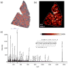
IMS analysis of a 109-year-old FFPE sample. (a) Contrast-enhanced Congo Red staining of a human spleen biopsy shows extensive amyloid deposits (in red) throughout the section. Scale bar, 5 mm. (b) MS image of a peptide at m/z 1550.7 from serum amyloid A localized to the areas of amyloid deposition. (c) MS/MS of this peptide directly from the tissue section resulted in nearly complete sequence coverage and identification of the peptide SFFSLGEAFDGAR.
Figure 4
Figure 4
H&E staining coupled with MS of an FFPE human kidney tumor. (a) A mass spectrum obtained from an H&E-stained section of a human kidney tumor. The inset shows the tissue that was analyzed, with 5× magnification of the area from which the spectrum was acquired. (b) The mass spectrum from an unstained serial section.

Similar articles

Cited by

References

    1. Andersson M, et al. Imaging mass spectrometry of proteins and peptides: 3D volume reconstruction. Nat Methods. 2008;5:101–108. - PubMed
    1. Burnum KE, et al. Imaging mass spectrometry reveals unique protein profiles during embryo implantation. Endocrinology. 2008;149:3274–3278. - PMC - PubMed
    1. Cazares LH, et al. Imaging mass spectrometry of a specific fragment of mitogen-activated protein kinase/extracellular signal-regulated kinase kinase kinase 2 discriminates cancer from uninvolved prostate tissue. Clin Cancer Res. 2009;15:5541–5551. - PubMed
    1. Lemaire R, et al. Direct Analysis and MALDI Imaging of Formalin-Fixed, Paraffin-Embedded Tissue Sections. Journal of Proteome Research. 2007;6:1295–1305. - PubMed
    1. Groseclose MR, et al. Identification of proteins directly from tissue: in situ tryptic digestions coupled with imaging mass spectrometry. J Mass Spectrom. 2007;42:254–262. - PubMed

Publication types

MeSH terms