. 2011 May;29(4):568-78.
doi: 10.1016/j.mri.2010.11.004. Epub 2011 Feb 2.

Beat-to-beat respiratory motion correction with near 100% efficiency: a quantitative assessment using high-resolution coronary artery imaging

Affiliations

Beat-to-beat respiratory motion correction with near 100% efficiency: a quantitative assessment using high-resolution coronary artery imaging

Andrew D Scott et al. Magn Reson Imaging. 2011 May.

Abstract

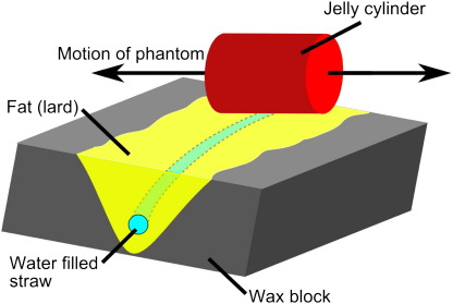
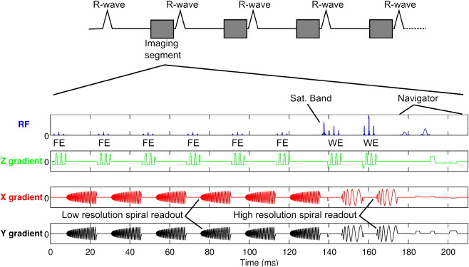
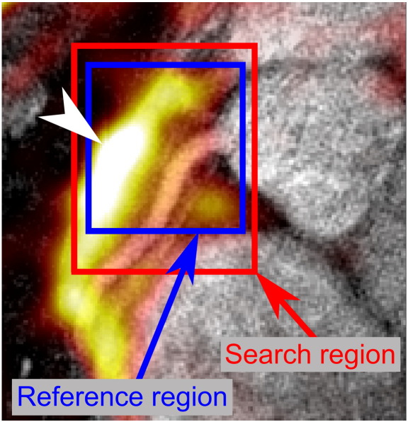
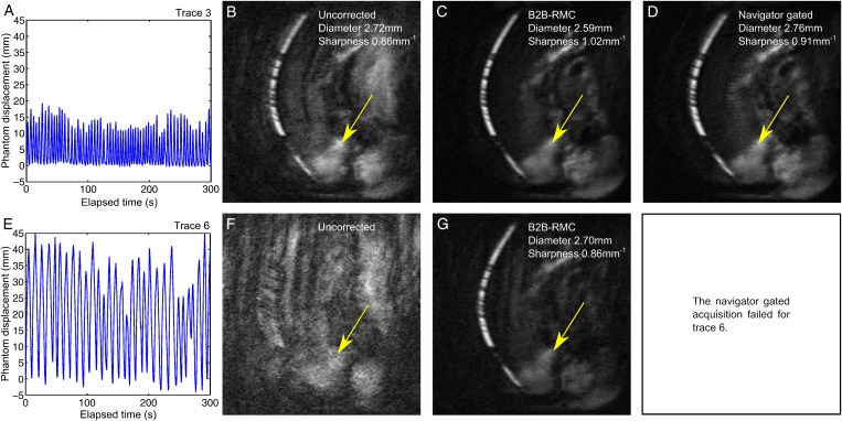
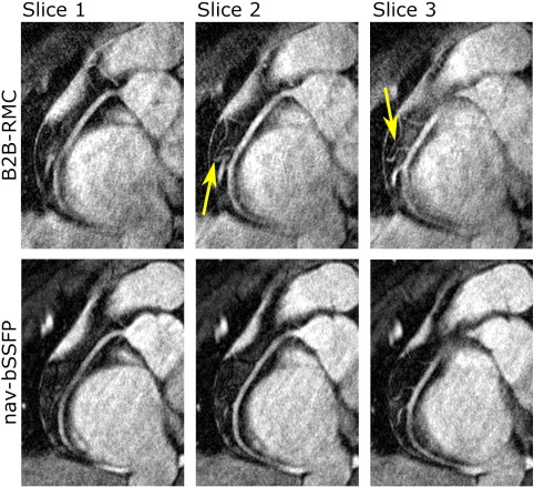
This study quantitatively assesses the effectiveness of retrospective beat-to-beat respiratory motion correction (B2B-RMC) at near 100% efficiency using high-resolution coronary artery imaging. Three-dimensional (3D) spiral images were obtained in a coronary respiratory motion phantom with B2B-RMC and navigator gating. In vivo, targeted 3D coronary imaging was performed in 10 healthy subjects using B2B-RMC spiral and navigator gated balanced steady-state free-precession (nav-bSSFP) techniques. Vessel diameter and sharpness in proximal and mid arteries were used as a measure of respiratory motion compensation effectiveness and compared between techniques. Phantom acquisitions with B2B-RMC were sharper than those acquired with navigator gating (B2B-RMC vs. navigator gating: 1.01±0.02 mm(-1) vs. 0.86±0.08 mm(-1), P<.05). In vivo B2B-RMC respiratory efficiency was significantly and substantially higher (99.7%±0.5%) than nav-bSSFP (44.0%±8.9%, P<.0001). Proximal and mid vessel sharpnesses were similar (B2B-RMC vs. nav-bSSFP, proximal: 1.00±0.14 mm(-1) vs. 1.08±0.11 mm(-1), mid: 1.01±0.11 mm(-1) vs. 1.05±0.12 mm(-1); both P=not significant [ns]). Mid vessel diameters were not significantly different (2.85±0.39 mm vs. 2.80±0.35 mm, P=ns), but proximal B2B-RMC diameters were slightly higher (2.85±0.38 mm vs. 2.70±0.34 mm, P<.05), possibly due to contrast differences. The respiratory efficiency of B2B-RMC is less variable and significantly higher than navigator gating. Phantom and in vivo vessel sharpness and diameter values suggest that respiratory motion compensation is equally effective.


Figures

Fig. 1
Fig. 1
The coronary artery test object consisted of a water-filled straw within a fat-filled groove in a wax block which was rotated with respect to the direction of motion. A jelly-filled cylinder was placed adjacent to the test object and orientated parallel to the direction of motion in order to monitor the phantom location with a standard navigator.
Fig. 2
Fig. 2
Sequence diagram for the B2B-RMC technique used for bright blood acquisition with 3D stack of spirals readouts. In every cardiac cycle, a full fat selective 3D low-resolution volume is acquired before the two interleaves from the high-resolution acquisition.
Fig. 3
Fig. 3
To aid positioning of the search and reference regions for the localized 3D cross-correlation of the fat images, the reference low-resolution fat volume was transparently overlaid (“hot” color scale - an arrow head indicates the center of the largest region of fat for clarity (in the black and white print version of this figure)) on the uncorrected high-resolution water images (“gray” scale). This enables accurate positioning of the regions around the right coronary origin which is otherwise challenging using the fat images alone. Both reference and search regions are defined in three dimensions, extending into multiple slices (not shown here for clarity).
Fig. 4
Fig. 4
Example results from the phantom tests using respiratory traces 3 (A) and 6 (E). Corresponding MIP images are shown from acquisitions without gating or correction (uncorrected) (B) and (F) and corrected using B2B-RMC (C) and (G). For respiratory trace 3, the MIP image from data acquired with a 5-mm navigator gating window is also shown (D). Respiratory efficiency of the uncorrected and B2B-RMC images (B, C, F, G) is 100% and 54% the navigator gated image (d). Some residual fat is seen in all of the images (arrows).
Fig. 5
Fig. 5
Example images from one subject showing corrected images from the B2B-RMC technique in three consecutive slices (top line) and the corresponding slices from the nav-bSSFP technique (bottom line). Note the similar appearance using both techniques of the right coronary artery in the proximal and midsections, but increased blurring in the B2B-RMC images in the distal vessel. Also note the improved depiction of the branch vessels in the midsection in the B2B-RMC images, highlighted by the arrows.
Fig. 6
Fig. 6
Curved planar reformat of 3D spiral images acquired with the B2B-RMC technique (0.7×0.7×3-mm resolution) in 302 cardiac cycles (99.3% efficient) over 20 mm of diaphragm motion, corrected for optimal proximal (A) and distal (B) vessel quality. For comparison, the equivalent curved planar reformat of the 3D nav-bSSFP with identical resolution acquired in 576 cardiac cycles (43.6% efficient) is shown (C).
Fig. 7
Fig. 7
The localized in-plane (bottom to top–y [i] and right to left–x [ii]) and through-plane (z [iii]) beat-to-beat displacements obtained from the low-resolution images using a cross-correlation technique around the (A) proximal and (B) distal artery, plotted against the diaphragm displacement obtained from the navigator. Inspiratory, expiratory and stationary points are differentiated using the difference between successive points in the navigator trace. The increased slope for the in-plane translations observed in the distal artery highlights the need for a localized correction.
Fig. 8
Fig. 8
Curved plane reformat of the data sets shown in Fig. 6A and B combined to correct for both proximal and distal motion. The data sets were combined in a basic manner, and further work will address more sophisticated methods.

References

    1. Danias P.G., McConnell M.V., Khasgiwala V.C., Chuang M.L., Edelman R.R., Manning W.J. Prospective navigator correction of image position for coronary MR angiography. Radiology. 1997;203(3):733–736. - PubMed
    1. Ehman R.L., Felmlee J.P. Adaptive technique for high-definition MR imaging of moving structures. Radiology. 1989;173(1):255–263. - PubMed
    1. Sachs T.S., Meyer C.H., Hu B.S., Kohli J., Nishimura D.G., Macovski A. Real-time motion detection in spiral MRI using navigators. Magn Reson Med. 1994;32(5):639–645. - PubMed
    1. Oshinski J.N., Hofland L., Mukundan S., Dixon W.T., Parks W.J., Pettigrew R.I. Two-dimensional coronary MR angiography without breath holding. Radiology. 1996;201(3):737–743. - PubMed
    1. Taylor A.M., Jhooti P., Wiesmann F., Keegan J., Firmin D.N., Pennell D.J. MR navigator-echo monitoring of temporal changes in diaphragm position: implications for MR coronary angiography. J Magn Reson Imaging. 1997;7(4):629–636. - PubMed

Publication types