. 2011 Nov;32(21):2713-22.
doi: 10.1093/eurheartj/ehq519. Epub 2011 Feb 2.

Fibroblast activation protein is induced by inflammation and degrades type I collagen in thin-cap fibroatheromata

Affiliations

Fibroblast activation protein is induced by inflammation and degrades type I collagen in thin-cap fibroatheromata

Chad E Brokopp et al. Eur Heart J. 2011 Nov.

Abstract

Aims: Collagen degradation in atherosclerotic plaques with thin fibrous caps renders them more prone to rupture. Fibroblast activation protein (FAP) plays a role in arthritis and tumour formation through its collagenase activity. However, the significance of FAP in thin-cap human fibroatheromata remains unknown.

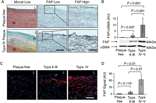
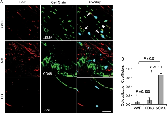
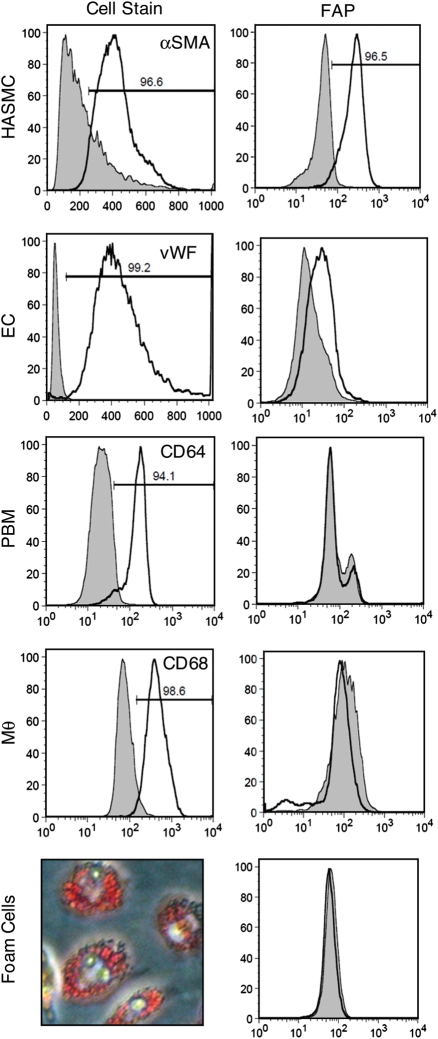
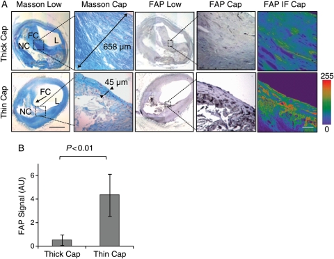
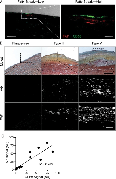
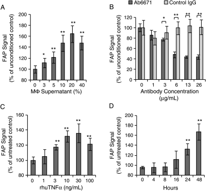
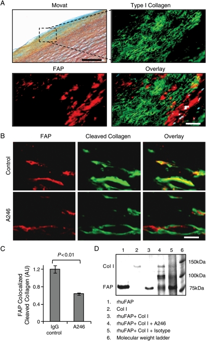
Methods and results: We detected enhanced FAP expression in type IV-V human aortic atheromata (n = 12), compared with type II-III lesions (n = 9; P < 0.01) and healthy aortae (n = 8; P < 0.01) by immunostaining and western blot analyses. Fibroblast activation protein was also increased in thin-cap (<65 µm) vs. thick-cap (≥ 65 µm) human coronary fibroatheromata (n = 12; P < 0.01). Fibroblast activation protein was expressed by human aortic smooth muscle cells (HASMC) as shown by colocalization on immunofluorescent aortic plaque stainings (n = 10; P < 0.01) and by flow cytometry in cell culture. Although macrophages did not express FAP, macrophage burden in human aortic plaques correlated with FAP expression (n = 12; R(2)= 0.763; P < 0.05). Enzyme-linked immunosorbent assays showed a time- and dose-dependent up-regulation of FAP in response to human tumour necrosis factor α (TNFα) in HASMC (n = 6; P < 0.01). Moreover, supernatants from peripheral blood-derived macrophages induced FAP expression in cultured HASMC (n = 6; P < 0.01), an effect abolished by blocking TNFα (n = 6; P < 0.01). Fibroblast activation protein associated with collagen-poor regions in human coronary fibrous caps and digested type I collagen and gelatin in vitro (n = 6; P < 0.01). Zymography revealed that FAP-mediated collagenase activity was neutralized by an antibody directed against the FAP catalytic domain both in HASMC (n = 6; P < 0.01) and in fibrous caps of atherosclerotic plaques (n = 10; P < 0.01).

Conclusion: Fibroblast activation protein expression in HASMC is induced by macrophage-derived TNFα. Fibroblast activation protein associates with thin-cap human coronary fibroatheromata and contributes to type I collagen breakdown in fibrous caps.


Figures

Figure 1
Figure 1
Fibroblast activation protein expression is enhanced in human atherosclerotic aortic plaques. (A) Movat and fibroblast activation protein stainings show cross-sections of representative plaque-free aortae and type IV aortic atherosclerotic plaques (L, lumen; M, media; P, atherosclerotic plaque; bar = 400 µm). Dotted boxes indicate regions of interest in adjacent sections at high magnification (bar = 50 µm). (B) Western blot analysis of fibroblast activation protein normalized to α-smooth muscle actin in plaque-free aortae (n = 8), type II–III plaques (n = 8), and type IV–V plaques (n = 7) shows a significant increase in fibroblast activation protein in advanced type IV–V plaques by immunoblot densitometry. (C) Immunofluorescent stainings in representative tissue sections of plaque-free aortae, type II–III plaque, and type IV plaque show fibroblast activation protein expression in red (DAPI in blue; bar = 50 µm). (D) The graph reveals a significant increase in fibroblast activation protein expression in type II–III aortic plaques (n = 9) and in type IV–V plaques (n = 12) compared with plaque-free aortae (n = 8).
Figure 2
Figure 2
Fibroblast activation protein expression in human aortic plaques colocalizes with smooth muscle cells, but not with macrophages or endothelial cells. (A) Overlays of confocal images of fibroblast activation protein (red) and DAPI (blue) with cell-specific stainings of α-smooth muscle actin, CD68, and von Willebrand factor (green) in representative sections illustrate fibroblast activation protein colocalization (arrows) with smooth muscle cells (bar = 20 µm). (B) The graph quantifies an increased colocalization of fibroblast activation protein with smooth muscle cells (α-smooth muscle actin), compared with endothelial cells (von Willebrand factor) and macrophages (CD68) in type IV–V atherosclerotic plaques (n = 10).
Figure 3
Figure 3
Fibroblast activation protein is constitutively expressed in cultured human aortic smooth muscle cells (HASMC) and human aortic endothelial cells (HAEC), but not in peripheral blood-derived monocytes (PBM), macrophages (MΦ), or foam cells. FACS analyses and Oil-Red-O staining of peripheral blood derived-macrophages laden with oxidized LDL characterize cells populations (left) and their respective fibroblast activation protein expression (right).
Figure 4
Figure 4
Fibroblast activation protein expression is enhanced in thin-cap vs. thick-cap human coronary fibroatheromata. (A) Masson staining shows collagen-rich thick (658 µm) vs. thin (45 µm) fibrous caps (L, lumen; FC, fibrous cap; NC, necrotic core; bar = 1 mm). Fibroblast activation protein immunohistochemistry and immunofluorescence (intensity scale; bar = 50 µm) shows fibroblast activation protein expression in representative thin vs. thick caps. Dotted boxes indicate regions of interest in adjacent sections at high magnification. (B) The graph reveals a significant increase in fibroblast activation protein expression in thin vs. thick fibrous caps (n = 12 each).
Figure 5
Figure 5
Fibroblast activation protein expression correlates with macrophage burden in human aortic plaques. (A) Confocal immunofluorescent photomicrograph of an aortic fatty streak reveals fibroblast activation protein expression (red) adjacent to macrophages (CD68; green) at low (phase-contrast, white; bar = 100 μm) and high magnification (bar = 25 μm). (B) Movat staining (bar = 400 μm), fibroblast activation protein, or macrophage (CD68) immunofluorescent stainings in plaque-free aortae, type II, and type V atherosclerotic plaques show enhanced fibroblast activation protein expression with increasing macrophage burden (bar = 50 µm). (C) Comparisons of fibroblast activation protein and macrophage expression in serial adjacent sections from aortic plaques demonstrate a significant positive correlation (R2= 0.763; n = 12; P < 0.05); AU, arbitrary units.
Figure 6
Figure 6
Macrophage-derived tumour necrosis factor α induces fibroblast activation protein expression in human aortic smooth muscle cells. (A) Macrophage-conditioned supernatant induces fibroblast activation protein in human aortic smooth muscle cells in a concentration-dependent manner following 48 h exposure (n = 6). (B) Using the same macrophage-conditioned medium, tumour necrosis factor α-blocking antibody (Ab6671) decreases fibroblast activation protein expression by 40% in human aortic smooth muscle cells compared with an isotype control antibody (n = 6). (C) Recombinant human tumour necrosis factor α induces fibroblast activation protein in human aortic smooth muscle cells in a dose-dependent manner after 48 h incubation (n = 6). (D) Recombinant human tumour necrosis factor α induces fibroblast activation protein in human aortic smooth muscle cells in a time-dependent manner (30 ng/mL). AU, arbitrary units (*P < 0.05, **P < 0.01).
Figure 7
Figure 7
Type I collagenase activity is inhibited in human aortic fibrous caps by the fibroblast activation protein-blocking antibody A246. (A) Movat staining of a fibrous cap in human aortic plaque. The region of interest (black box) is shown at higher magnification in an adjacent section stained for fibroblast activation protein (red), type I collagen (green), and overlay (DAPI = blue; bar = 150 µm). (B) Confocal images of in situ zymography show fibroblast activation protein (red) and cleaved DQ type I collagen (green) in fibrous caps shown by Movat (A) of aortic plaque treated with a control IgG antibody or neutralizing antibody A246 (bar = 10 μm). (C) The graph reveals a significant reduction of cleaved type I collagen colocalized with fibroblast activation protein expression by in situ zymography (n = 10/group). (D) Type I collagenase activity of recombinant human fibroblast activation protein and the neutralizing capacity of A246 was demonstrated by incubation of native human type I collagen with recombinant human fibroblast activation protein in the presence of inhibiting or isotype control antibodies (lanes: 1, rhuFAP; 2, collagen; 3, rhuFAP + collagen; 4, rhuFAP + collagen + A246; 5, rhuFAP + collagen + isotype control antibody; 6, molecular weight marker).

References

    1. Farb A, Burke AP, Tang AL, Liang Y, Mannan P, Smialek J, Virmani R. Coronary plaque erosion without rupture into a lipid core: a frequent cause of coronary thrombosis in sudden coronary death. Circulation. 1996;93:1354–1363. - PubMed
    1. Virmani R, Burke AP, Farb A, Kolodgie FD. Pathology of the vulnerable plaque. J Am Coll Cardiol. 2006;47:C13–C18. doi:10.1016/j.jacc.2005.10.065. - DOI - PubMed
    1. van der Wal A, Becker A, van der Loos C, Das P. Site of intimal rupture or erosion of thrombosed coronary atherosclerotic plaques is characterized by an inflammatory process irrespective of the dominant plaque morphology. Circulation. 1994;89:36–44. - PubMed
    1. Shah P, Falk E, Badimon J, Fernandez-Ortiz A, Mailhac A, Villareal-Levy G, Fallon J, Regnstrom J, Fuster V. Human monocyte-derived macrophages induce collagen breakdown in fibrous caps of atherosclerotic plaques. Potential role of matrix-degrading metalloproteinases and implications for plaque rupture. Circulation. 1995;92:1565–1569. - PubMed
    1. Rajavashisth TB, Xu X-P, Jovinge S, Meisel S, Xu X-O, Chai N-N, Fishbein MC, Kaul S, Cercek B, Sharifi B, Shah PK. Membrane type 1 matrix metalloproteinase expression in human atherosclerotic plaques. Circulation. 1999;99:3103–3109. - PubMed

Publication types

MeSH terms