. 2011 Mar;13(3):267-79.
doi: 10.1093/neuonc/noq195. Epub 2011 Feb 3.

Amiodarone sensitizes human glioma cells but not astrocytes to TRAIL-induced apoptosis via CHOP-mediated DR5 upregulation

Affiliations

Amiodarone sensitizes human glioma cells but not astrocytes to TRAIL-induced apoptosis via CHOP-mediated DR5 upregulation

In Young Kim et al. Neuro Oncol. 2011 Mar.

Abstract

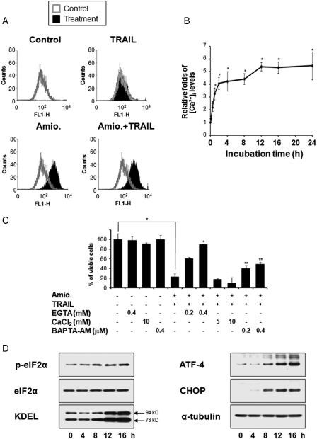
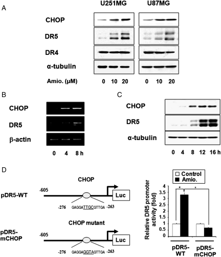
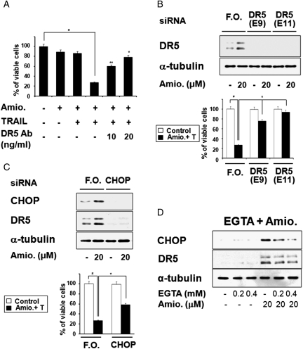
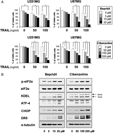
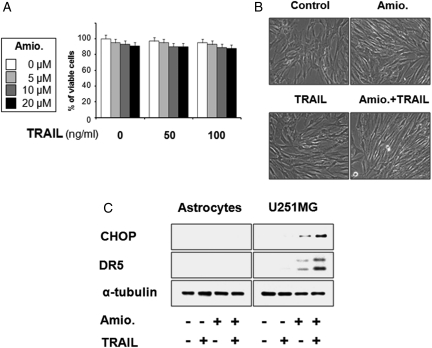
Amiodarone is a widely used anti-arrhythmic drug that inhibits diverse ion channels, including the Na(+)/Ca(2+) exchanger (NCX), L-type Ca(2+) channels, and Na(+) channels. Here, we report that subtoxic doses of amiodarone and tumor necrosis factor-related apoptosis-inducing ligand (TRAIL) synergistically induced apoptosis of various glioma cells. Treatment of U251MG glioma cells with amiodarone increased intracellular Ca(2+) levels and enhanced the expression of the endoplasmic reticulum (ER) stress-inducible transcription factor C/EBP homologous protein (CHOP). This upregulation of CHOP was followed by marked upregulation of the TRAIL receptor, DR5. Suppression of DR5 expression by small interfering (si) RNAs almost completely blocked amiodarone/TRAIL-induced apoptosis in U251MG glioma cells, demonstrating that DR5 is critical to this cell death. siRNA-mediated CHOP suppression reduced amiodarone-induced DR5 upregulation and attenuated the cell death induced by amiodarone plus TRAIL. In addition, omitting Ca(2+) from the external medium using ethylene glycol tetraacetic acid markedly inhibited this cell death, reducing the protein levels of CHOP and DR5. These results suggest that amiodarone-induced influx of Ca(2+) plays an important role in sensitizing U251MG cells to TRAIL-mediated apoptosis through CHOP-mediated DR5 upregulation. Furthermore, subtoxic doses of bepridil and cibenzoline, two other anti-arrhythmic drugs with NCX-inhibitor activity, also sensitized glioma cells to TRAIL-mediated apoptosis, via the upregulation of both CHOP and DR5. Notably, amiodarone/TRAIL cotreatment did not induce cell death in astrocytes, nor did it affect the expression of CHOP or DR5 in these cells. These results collectively suggest that a combined regimen of amiodarone plus TRAIL may offer an effective therapeutic strategy for safely and selectively treating resistant gliomas.


Figures

Fig. 1.
Fig. 1.
Combined treatment with amiodarone and TRAIL effectively induces TRAIL-mediated apoptosis in glioma cells. (A) Effect of amiodarone and/or TRAIL on the viability of glioma cells. U251MG, U87MG, U343, and U251N cells were treated with amiodarone for 30 min and then further treated with TRAIL for 24 h at the indicated concentrations. Cellular viability was assessed using calcein-AM and EthD-1. Columns indicate average of 3 individual experiments; bars represents ±SD; #P < 0.01, compared with untreated cells; *P < 0.001, compared with TRAIL-treated cells; **P < 0.01, compared with TRAIL-treated cells. (B) Synergistic induction of cell death by amiodarone and TRAIL. Four different glioma cells were treated for 24 h with increasing concentrations of amiodarone and TRAIL. Isobologram analysis was performed as described in Materials and Methods. IC50 for amiodarone or TRAIL in the respective glioma cells are as follows: U251MG (50 µM, 500 ng/ml); U87MG (90 µM, 360 ng/ml); U343 (85 µM, 40 ng/ml); U251N (220 µM, 93 ng/ml). (C) Activation of caspases following combined treatment with amiodarone and TRAIL. U251MG cells were treated with 20 µM amiodarone (Amio.) alone, 100 ng/ml TRAIL (T) alone, or amiodarone plus TRAIL (Amio. + T) for the indicated time points. Western blotting of the caspases, PARP, Bid, and α-tubulin was performed. (D) Effect of caspases on the cell death induced by amiodarone and TRAIL. U251MG cells were treated with the respective caspase-specific tetrapeptide inhibitors at the indicated concentrations for 30 min and further treated with 20 µM amiodarone and 100 ng/ml TRAIL for 24 h. Cellular viability was determined using calcein-AM and EhtD-1. The graph represents the result of 1 of 3 independent experiments with consistent results. Columns indicate average of 3 individual experiments; bars represents ±SD; #P < 0.001, compared with untreated cells; *P < 0.001, compared with cells treated with TRAIL alone; **P < 0.01, compared with cells treated with TRAIL alone.
Fig. 2.
Fig. 2.
Amiodarone induces ER stress. (A) Effect of amiodarone and/or TRAIL on the intracellular Ca2+ levels. U251MG cells were treated with 20 µM amiodarone alone, 100 ng/ml TRAIL alone, or amiodarone plus TRAIL for 24 h. Flow cytometry using fluo-3 was performed as described in Materials and Methods. (B) Changes in the intracellular Ca2+ levels in response to amiodarone. U251MG cells were treated with 20 µM amiodarone for the indicated time points, analyzed by flow cytometry. Dots indicate average of 3 individual experiments; bars represents ±SD; *P < 0.001, compared with untreated cells. (C) Effect of EGTA, CaCl2, or BAPTA-AM on the cell death induced by amiodarone plus TRAIL. U251MG cells were treated with EGTA, CaCl2, or BAPTA-AM at the indicated concentrations and further treated with 20 µM amiodarone and 100 ng/ml TRAIL for 24 h. Cellular viability was determined using calcein-AM and EhtD-1. The graph represents the result of 1 of 3 independent experiments with consistent results. Columns indicate average of 3 individual experiments; bars represents ±SD; #P < 0.001, compared with untreated cells; *P < 0.001, compared with amiodarone plus TRAIL-treated cells; **P < 0.01, compared with amiodarone plus TRAIL-treated cells. (D) Changes in the protein levels associated with ER stress following amiodarone treatment. U251MG cells were treated with 20 µM amiodarone for the indicated times and Western blotting was performed. Western blotting of α-tubulin served as a loading control of the samples.
Fig. 3.
Fig. 3.
CHOP-mediated DR5 upregulation is critical for amidarone-stimulated TRAIL-induced apoptosis. (A) Amiodarone increased the protein levels of CHOP and DR5. U251MG and U87MG cells were treated with amiodarone at the indicated concentrations for 24 h, and Western blotting of CHOP, DR5, DR4, and α-tubulin was performed. (B) Changes in the mRNA levels of CHOP and DR5 following amiodarone treatment. U251MG cells were treated with 20 µM amiodarone at the indicated time points, and RT-PCR was performed to detect the mRNA levels of CHOP, DR5, and β-actin. (C) Changes in the protein levels of CHOP and DR5 following amiodarone treatment. U251MG cells were treated with 20 µM amiodarone at the indicated time points, and Western blotting of CHOP and DR5 was performed. Western blotting of α-tubulin served as a loading control. (D) Mutation at CHOP binding site of the DR5 promoter abolishes amiodarone-mediated transcriptional activation of DR5. U251MG cells were transfected with pDR5-WT or CHOP-mutated pDR5-mCHOP and then treated with 20 µM amiodarone for 12 h, lysed, and assayed for luciferase activity. Columns indicate average of 3 individual experiments; bars represents ±SD; #P < 0.001, compared with untreated cells; *P < 0.001, compared with cell transfected with pDR5-WT and further treated with amiodarone.
Fig. 4.
Fig. 4.
CHOP-mediated DR5 upregulation contributes to amiodarone-stimulated TRAIL-mediated apoptosis. (A) Effect of DR5-specific blocking chimera antibody on amiodarone/TRAIL-induced apoptosis. U251MG cells were pretreated with or without amiodarone for 30 min, followed by treatment with or without 100 ng/ml TRAIL for 24 h in the presence of indicated concentrations of DR5-specific blocking chimera antibody. Cellular viabilities were measured with calcein-AM and EthD-1 to detect live and dead cells, respectively. Similar results were obtained from 3 independent experiments; bars represent ±SD; #P < 0.001, compared with untreated cells; *P < 0.001, compared with amiodarone plus TRAIL–treated cells; **P < 0.01, compared with amiodarone plus TRAIL–treated cells. (B) Suppression of DR5 expression by siRNA blocks amiodarone-stimulated TRAIL-induced apoptosis in U251MG cells. U251MG cells were transfected with the control fluorescent oligonucleotide (F.O.) or two different siRNA duplexes against DR5 mRNA (E9 and E11). Twenty-four hours after transfection, cells were further treated with or without 20 µM amiodarone for 24 h. Western blotting of DR5 was done to confirm the downregulation of DR5 by siRNA transfection. α-Tubulin levels were assessed to show equal gel loading. To examine the effect of DR5 downregulation on amiodarone/TRAIL-induced apoptosis, U251MG cells were transfected with siRNAs, incubated for 24 h, and further treated with or without 20 µM amiodarone plus 100 ng/ml TRAIL for 24 h. Cellular viability was determined using calcein-AM and EthD-1. Columns indicate average of 3 individual experiments; bars represent ±SD; #P < 0.01, compared with untreated cells; *P < 0.01, compared with the cells transfected with the F.O. and further treated with amiodarone plus TRAIL. (C) Role of CHOP in amiodarone-induced DR5 upregulation and amiodarone-stimulated TRAIL-induced apoptosis. Suppression of CHOP expression by siRNA reduces amiodarone-induced DR5 upregulation and amiodarone-stimulated TRAIL-induced apoptosis in U251MG cells. U251MG cells were transfected with the control F.O. or siRNA duplexes against CHOP, incubated for 24 h, and further treated with 20 µM amiodarone alone for 24 h. First, Western blotting of CHOP was performed to confirm the downregulation of CHOP by siRNA transfection. Western blotting of DR5 was also performed to examine the knockdown effect of CHOP on amiodarone-induced DR5 upregulation. Equal loading of the protein samples was confirmed by Western blotting of α-tubulin. To examine the effect of CHOP downregulation on amiodarone-sensitized TRAIL-induced apoptosis, transfected cells with the control F.O. or CHOP siRNA were treated with 20 µM amiodarone plus 100 ng/ml TRAIL for 24 h. Cellular viability was determined using calcein-AM and EthD-1. Columns indicate average of 3 individual experiments; bars represent ±SD; #P < 0.01, compared with untreated cells; * P < 0.01, compared with the cells transfected with the F.O and further treated with amiodarone plus TRAIL. (D) Effect of EGTA on amiodarone-induced upregulation of CHOP and DR5. U251MG cells were pretreated with EGTA at the indicated concentrations and further treated with 20 µM amiodarone for 24 h. Western blotting of CHOP, DR5, and α-tubulin was performed.
Fig. 5.
Fig. 5.
The anti-arrythmic drugs bepridil and cibenzoline also sensitize glioma cells to TRAIL-mediated apoptosis via CHOP-mediated DR5 upregulation. (A) Effect of bepridil plus TRAIL or cibenzoline plus TRAIL on the viability of various glioma cells. U251MG and U87MG cells were treated with bepridil or cibenzoline for 30 min and then further treated with TRAIL for 24 h at the indicated concentrations. Cellular viability was assessed using calcein-AM and EthD-1. Columns indicate average of 3 individual experiments; bars represent ± SD; #P < 0.001, compared with untreated cells; *P < 0.001, compared with TRAIL-treated cells. (B) Effect of bepridil or cibenzoline on the expression of ER stress–associated proteins and DR5. U251MG cells were treated with bepridil or cibenzoline for 24 h at the indicated concentrations. Western blotting to detect changes in the expression of indicated proteins was performed.
Fig. 6.
Fig. 6.
Effect of amiodarone and/or TRAIL on the viability of human normal astrocytes. (A) Human astrocytes were treated with amdiodarone and/or TRAIL at the indicated concentrations for 24 h. Cellular viability was assessed using calcein-AM and EthD-1. (B) Human astrocytes were treated with 20 µM amiodarone for 30 min and further treated with or without 100 ng/ml TRAIL for 24 h. Morphologies of human astrocytes treated with amiodarone and/or TRAIL are shown. (C) Effect of amiodarone/or TRAIL on the expression of CHOP and DR5 in astrocytes. Astrocytes and U251MG cells were treated with 20 µM amiodarone alone, 100 ng/ml TRAIL alone, or a combination of both for 24 h, and cell extracts were prepared for Western blotting of CHOP and DR5. Western blotting of α-tubulin served as a loading control of the sample.

Similar articles

Cited by

References

    1. Ashkenazi A, Dixit VM. Apoptosis control by death and decoy receptors. Curr Opin Cell Biol. 1999;11(2):255–260. - PubMed
    1. Hawkins CJ. TRAIL and malignant glioma. Vitam Horm. 2004;67:427–452. - PubMed
    1. Kleihues P, Cavenee WK. World Health Organization Classification of Tumors. Pathology and Genetics: Tumors of the Nervous System. 2nd. Albany, NY: WHO Publication Center USA; 2000.
    1. Maher EA, Furnari FB, Bachoo RM, et al. Malignant glioma: genetics and biology of a grave matter. Genes Dev. 2001;15(11):1311–1333. - PubMed
    1. Knight MJ, Riggkin CD, Muscat AM, et al. Analysis of FasL and TRAIL induced apoptosis pathways in glioma cells. Oncogene. 2001;20(41):5789–5798. - PubMed

Publication types

MeSH terms