. 2011 Apr 8;286(14):12724-33.
doi: 10.1074/jbc.M110.177089. Epub 2011 Feb 3.

Modulating micro-opioid receptor phosphorylation switches agonist-dependent signaling as reflected in PKCepsilon activation and dendritic spine stability

Affiliations

Modulating micro-opioid receptor phosphorylation switches agonist-dependent signaling as reflected in PKCepsilon activation and dendritic spine stability

Hui Zheng et al. J Biol Chem. .

Abstract

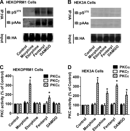
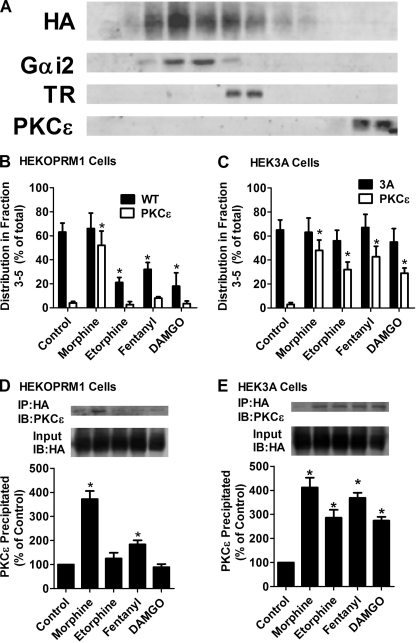
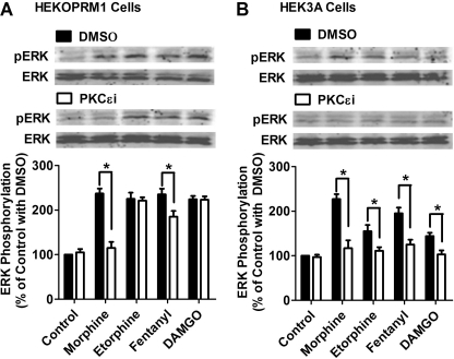
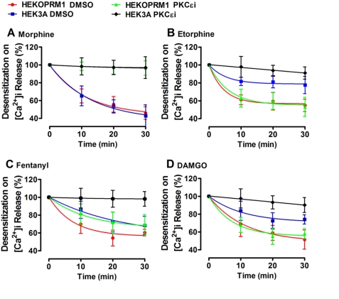
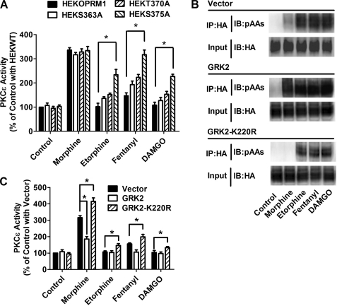
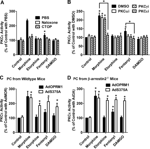
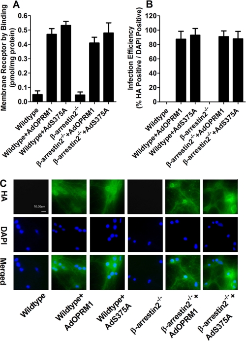
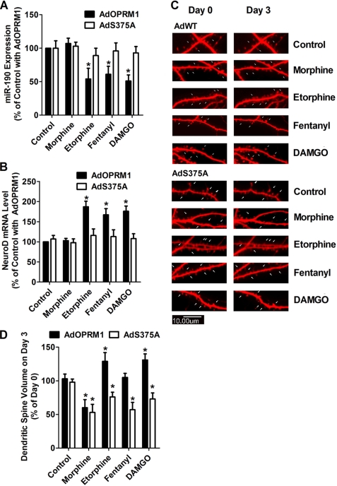
A new role of G protein-coupled receptor (GPCR) phosphorylation was demonstrated in the current studies by using the μ-opioid receptor (OPRM1) as a model. Morphine induces a low level of receptor phosphorylation and uses the PKCε pathway to induce ERK phosphorylation and receptor desensitization, whereas etorphine, fentanyl, and [D-Ala2,N-Me-Phe4,Gly5-ol]-enkephalin (DAMGO) induce extensive receptor phosphorylation and use the β-arrestin2 pathway. Blocking OPRM1 phosphorylation (by mutating Ser363, Thr370 and Ser375 to Ala) enabled etorphine, fentanyl, and DAMGO to use the PKCε pathway. This was not due to the decreased recruitment of β-arrestin2 to the receptor signaling complex, because these agonists were unable to use the PKCε pathway when β-arrestin2 was absent. In addition, overexpressing G protein-coupled receptor kinase 2 (GRK2) decreased the ability of morphine to activate PKCε, whereas overexpressing dominant-negative GRK2 enabled etorphine, fentanyl, and DAMGO to activate PKCε. Furthermore, by overexpressing wild-type OPRM1 and a phosphorylation-deficient mutant in primary cultures of hippocampal neurons, we demonstrated that receptor phosphorylation contributes to the differential effects of agonists on dendritic spine stability. Phosphorylation blockage made etorphine, fentanyl, and DAMGO function as morphine in the primary cultures. Therefore, agonist-dependent phosphorylation of GPCR regulates the activation of the PKC pathway and the subsequent responses.


Figures

FIGURE 1.
FIGURE 1.
Receptor phosphorylation attenuates PKCϵ activation. HEKOPRM1 (A and C) and HEK3A cells (B and D) were treated with PBS (Control), 1 μm morphine, 10 nm etorphine, 10 nm fentanyl and 1 μm DAMGO for 5 min. A and B, receptors were immunoprecipitated with HA antibody. The phosphorylated Ser375 on OPRM1 (pS375) and phosphorylated amino acid (pAAs) were determined in the immunoprecipitated receptor. C and D, activities of three PKC subtypes were determined as described under “Experimental Procedures.” The results were normalized against that in the control in each group.
FIGURE 2.
FIGURE 2.
Receptor phosphorylation attenuates the recruitment of PKCϵ. A, distributions of OPRM1, Gαi2, transferrin receptor (TR), and PKCϵ in continuous sucrose gradient in untreated HEKOPRM1 cells. B–E, HEKOPRM1 (A and C) and HEK3A cells (B and D) were treated as in Fig. 1. B and C, cells were subjected to continuous sucrose gradient and immunoblotting as described. The amounts of receptor and PKCϵ from fraction 3 to fraction 5 were normalized to the total amounts (from fraction 1 to fraction 12). C and D, receptors were immunoprecipitated with HA antibody. The amounts of PKCϵ co-immunoprecipitated with receptor were determined. The results were normalized against that in the control.
FIGURE 3.
FIGURE 3.
Phosphorylation blockage enables agonists to use PKCϵ for ERK phosphorylation. Cells were treated with DMSO or 50 μm PKCϵ-specific inhibitor (PKCϵi) for 3 h. HEKOPRM1 (A) and HEK3A cells (B) were then treated with agonists as in Fig. 1. ERK phosphorylation was determined by normalizing the immunoreactivities of phosphorylated ERK against the immunoreactivities of total ERK. The results were further normalized against that in the control with DMSO.
FIGURE 4.
FIGURE 4.
Phosphorylation blockage enables agonists to use PKCϵ for receptor desensitization. HEKOPRM1 and HEK3A were treated with DMSO or 50 μm PKCϵi for 3 h and were then pretreated with 100 nm morphine (A), 1 nm etorphine (B), 1 nm fentanyl (C), and 100 nm DAMGO (D) for the times indicated on the x axis. ADP was added after agonist pretreatment. The percentage decrease in the agonist-induced potentiation on the ADP-induced [Ca2+]i release was used to indicate the receptor desensitization. The decreases were calculated by using the formula: 100% − potentiation on ADP-induced [Ca2+]i release with pretreatment/potentiation on ADP-induced [Ca2+]i release without pretreatment.
FIGURE 5.
FIGURE 5.
GRK2 phosphorylation on Ser375 attenuates PKCϵ activation. A, HEKOPRM1, HEKS363A, HEKT370A, and HEKS375A cells were treated with PBS (Control), 1 μm morphine, 10 nm etorphine, 10 nm fentanyl, and 1 μm DAMGO for 5min. The activity of PKCϵ was determined. B and C, HEKOPRM1 was transfected with a vector, GRK2, and GRK2-K220R. One day after transfection, cells were treated with agonists as in A. Receptor phosphorylation on Ser375 was determined in B. PKCϵ activities were normalized against that in “Control with Vector” in C.
FIGURE 6.
FIGURE 6.
Receptor phosphorylation attenuates PKCϵ activation in primary cultures. A and B, primary cultures of hippocampal neurons from wild-type mice were pretreated with PBS, 10 μm naloxone, or 10 μm CTOP for 10 min (A). The cultures were pretreated with DMSO or 50 μm PKC subtype-specific inhibitors (PKCαi, PKCγI, and PKCϵi) for 3 h (B). Then the primary cultures were incubated with 1 μm morphine, 10 nm etorphine, 10 nm fentanyl, or 1 μm DAMGO for 5 min, and the activities of PKC subtypes were determined as described under “Experimental Procedures.” The results were normalized against that of the control with PBS (A) and the control with DMSO (B). C, primary hippocampal neurons from wild-type mice were infected with AdOPRM1 or AdS375A for 3 days. Then the primary cultures were incubated with 1 μm morphine, 10 nm etorphine, 10 nm fentanyl, or 1 μm DAMGO for 5 min, and the activity of PKCϵ was determined. The results were normalized against that of the control with AdOPRM1. D, primary hippocampal neurons from βarrestin2−/− mice were prepared as in C, and PKCϵ activity was determined.
FIGURE 7.
FIGURE 7.
High efficiency of virus infection leads to high receptor expression. Primary cultures of hippocampal neurons from wild-type or β-arrestin2−/− mice were infected with AdOPRM1 or AdS375A for 3 days. The receptor expression was determined with binding assay after infection (A). The efficiency of the infection was indicated by the percentage of HA-positive (green in C) cells in DAPI-positive (blue in C) cells (B and C).
FIGURE 8.
FIGURE 8.
Phosphorylation blockage switches the effects of agonist on spine stability. A and B, primary hippocampal neurons from wild-type mice were infected with AdOPRM1 or AdS373A for 3 days. Then the primary cultures were incubated with 1 μm morphine, 10 nm etorphine, 10 nm fentanyl, or 1 μm DAMGO for additional 3 days. The levels of miR-190 (A) and NeuroD mRNA (B) were determined. The results were normalized against that of the control with AdOPRM1. C and D, primary hippocampal neurons from wild-type mice were transfected with DsRed in pRK5 7 days after plating. Two weeks later, the cultures were infected with AdOPRM1 or AdS375A for 3 days. Then the primary cultures were incubated with 1 μm morphine, 10 nm etorphine, 10 nm fentanyl, or 1 μm DAMGO for additional 3 days. At Day 0 and Day 3 of agonist treatment, dendritic spine stability was examined in confocal images as described under “Experimental Procedures.” The spine densities (spine volume) on Day 3 were normalized against that on Day 0 and summarized (C). The images indicate the changes on spine morphology (D).

Similar articles

Cited by

References

    1. Urban J. D., Clarke W. P., von Zastrow M., Nichols D. E., Kobilka B., Weinstein H., Javitch J. A., Roth B. L., Christopoulos A., Sexton P. M., Miller K. J., Spedding M., Mailman R. B. (2007) J. Pharmacol. Exp. Ther. 320, 1–13 - PubMed
    1. Violin J. D., Lefkowitz R. J. (2007) Trends Pharmacol. Sci. 28, 416–422 - PubMed
    1. DeWire S. M., Ahn S., Lefkowitz R. J., Shenoy S. K. (2007) Annu. Rev. Physiol. 69, 483–510 - PubMed
    1. Zheng H., Loh H. H., Law P. Y. (2008) Mol. Pharmacol. 73, 178–190 - PMC - PubMed
    1. Kobayashi H., Narita Y., Nishida M., Kurose H. (2005) Cell Signal. 17, 1248–1253 - PubMed

Publication types