. 2011 Apr 8;286(14):12495-508.
doi: 10.1074/jbc.M110.209130. Epub 2011 Feb 3.

The low resolution structure of ApoA1 in spherical high density lipoprotein revealed by small angle neutron scattering

Affiliations

The low resolution structure of ApoA1 in spherical high density lipoprotein revealed by small angle neutron scattering

Zhiping Wu et al. J Biol Chem. .

Abstract

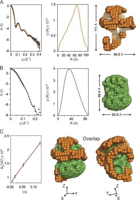
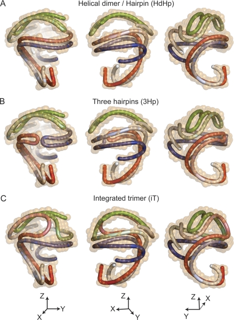
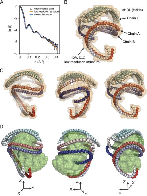
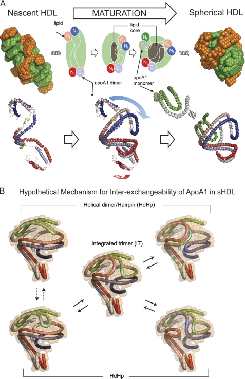
Spherical high density lipoprotein (sHDL), a key player in reverse cholesterol transport and the most abundant form of HDL, is associated with cardiovascular diseases. Small angle neutron scattering with contrast variation was used to determine the solution structure of protein and lipid components of reconstituted sHDL. Apolipoprotein A1, the major protein of sHDL, forms a hollow structure that cradles a central compact lipid core. Three apoA1 chains are arranged within the low resolution structure of the protein component as one of three possible global architectures: (i) a helical dimer with a hairpin (HdHp), (ii) three hairpins (3Hp), or (iii) an integrated trimer (iT) in which the three apoA1 monomers mutually associate over a portion of the sHDL surface. Cross-linking and mass spectrometry analyses help to discriminate among the three molecular models and are most consistent with the HdHp overall architecture of apoA1 within sHDL.


Figures

FIGURE 1.
FIGURE 1.
Small angle neutron scattering results. A, neutron scattering results for sHDL in 12% D2O. Left panel, the scattering intensity as a function of the scattering vector q. The experimental data are plotted with open circles and have error bars attached. The experimental scattering curve displays oscillations with maxima at ∼0.1, 0.2, and 0.25 Å−1. The solid orange line is the scattering intensity calculated from the low resolution structure obtained by deconvoluting the experimental data. Middle panel, the distance distribution function, p(R), plotted as a function of the distance between scattering centers. The p(R) value was obtained by deconvoluting the experimental scattering intensities of sHDL in 12% D2O with the program GNOM. Right panel, low resolution structure of the protein component of sHDL obtained from the p(R) function (middle panel) with the program DAMMIN. B, neutron scattering results for sHDL in 42% D2O. Left panel, the scattering intensity (open circles) for sHDL in 42% D2O. The error bars are very small and only slightly visible at the center of the open circles. The solid green line is the scattering intensity produced by the low resolution structure of the lipid. Middle panel, the p(R) function for the lipid component of sHDL. Right panel, low resolution structure of the lipid component of sHDL obtained from the p(R) function (middle panel). C, left panel, the Stuhrmann plot gives the dependence of Rg2 (where Rg is radius of gyration) on the contrast level (X) in the SANS experiment. A linear graph (red line) indicates that the protein component is located on the outside, and the lipid component is inside as shown by the overlap of the low resolutions structures for the 12% D2O (orange) and 42% D2O (green) in the middle and right panels. X, Y, and Z represent the directions of the Cartesian coordinate system.
FIGURE 2.
FIGURE 2.
Possible architectures of the apoA1 trimer in spherical HDL. A, superposition of the 12% D2O low resolution structure of sHDL (orange) with a schematic representation of the apoA1 trimer made of a helical dimer (red/blue) and a hairpin (green): the HdHp model. The three chains are gradient-colored (dark N terminus and lighter colored C terminus). The three panels depict different orientations of the HdHp model. B, the architecture of the apoA1 trimer corresponds to three hairpins that fit the 12% D2O low resolution shape: the 3Hp model. The red and blue hairpins interact with their helix 5 domains. The green hairpin retains the same orientation as the one in A. C, in this architecture (iT model) the apoA1 trimer has each chain associated with each other one, producing three dimers. All chains interact together with their helix 5 domains.
FIGURE 3.
FIGURE 3.
Comparison of experimental neutron scattering intensities with those obtained from the low resolution structure and the molecular model of the apoA1 trimer. A, the fit of scattering intensities produced by the 12% D2O low resolution structure (orange curve) and the molecular model (blue curve) of the apoA1 trimer (HdHp model) to the experimental data. B, the superposition of the molecular model of the apoA1 trimer (HdHp) with the 12% D2O low resolution structure (semitransparent orange beads). The molecular model of the apoA1 trimer is shown as three chains, two of them in a helical dimer conformation, and the third chain forms a hairpin. Because the molecular model does not include any local information about the conformation of the individual amino acid residues, the secondary structure of the chains was assigned to be α-helix for all residues. The three chains are gradient-colored red, blue and cyan. C and D, superposition of the HdHp model with the low resolution structure (orange) of the protein component (12% D2O) of sHDL (C) and the low resolution structure of the lipid component (42% D2O) (D).
FIGURE 4.
FIGURE 4.
Hypothetical mechanism of conformational change of apoA1 in nascent HDL during maturation into spherical HDL. A, left, superposition of the low resolution structures of protein (orange) and lipid (green) components of nascent HDL (top) and one particular orientation of the double super helix model of the protein component in nascent HDL (bottom left). The two apoA1 chains are gradient-colored red and blue, and the putative lecithin:cholesterol acyltransferase-binding loops (solar flares) are colored yellow. Middle, overlap of the middle domains of nascent HDL and sHDL apoA1 dimers. The overlapping suggests how the N and C termini of the apoA1 dimer in nascent HDL might swing and rearrange during particle maturation into what is found to be the dimer conformation in sHDL. The gray region at the middle of the particle represents the growing core of neutral lipids that accumulate during maturation. Right, superposition of the low resolution structures of protein (orange) and lipid (green) components of spherical HDL (top), and the resultant architecture of apoA1 trimer in spherical HDL (HdHp model, bottom right). B, hypothetical mechanism for apoA1 inter-exchange in sHDL. The hairpin and the apoA1 dimer are shown in different conformations that match the low resolution structure (12% D2O) of the protein. The different configurations suggest that it is reasonable to expect that because of particle dynamics the hairpin can bend such that it aligns its h5 domain with the h5 domain of the dimer (dotted double arrow lines) as a preamble for annealing with the dimer. Thus, the hairpin can exchange with one of the apoA1 monomers of the dimer through a transient integrated trimer-like configuration (center), generating other arrangements of the helical dimer/hairpin combination (solid double arrow lines). This protein reorganization mechanism makes all apoA1 monomers equivalent from an exchange point of view and can, in principle, lead to the integrated trimer (iT model) as illustrated on the right.

Similar articles

Cited by

References

    1. Castelli W. P., Doyle J. T., Gordon T., Hames C. G., Hjortland M. C., Hulley S. B., Kagan A., Zukel W. J. (1977) Circulation 55, 767–772 - PubMed
    1. Durrington P. N., Ishola M., Hunt L., Arrol S., Bhatnagar D. (1988) Lancet 1, 1070–1073 - PubMed
    1. Pekkanen J., Linn S., Heiss G., Suchindran C. M., Leon A., Rifkind B. M., Tyroler H. A. (1990) New Engl. J. Med. 322, 1700–1707 - PubMed
    1. Karathanasis S. K., Norum R. A., Zannis V. I., Breslow J. L. (1983) Nature 301, 718–720 - PubMed
    1. Norum R. A., Lakier J. B., Goldstein S., Angel A., Goldberg R. B., Block W. D., Noffze D. K., Dolphin P. J., Edelglass J., Bogorad D. D., Alaupovic P. (1982) New Engl. J. Med. 306, 1513–1519 - PubMed

Publication types

LinkOut - more resources