. 2011 Apr 8;286(14):11970-6.
doi: 10.1074/jbc.M110.190215. Epub 2011 Feb 3.

Identification of essential lysines involved in substrate binding of vacuolar H+-pyrophosphatase

Affiliations

Identification of essential lysines involved in substrate binding of vacuolar H+-pyrophosphatase

Chien-Hsien Lee et al. J Biol Chem. .

Abstract

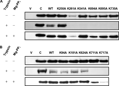
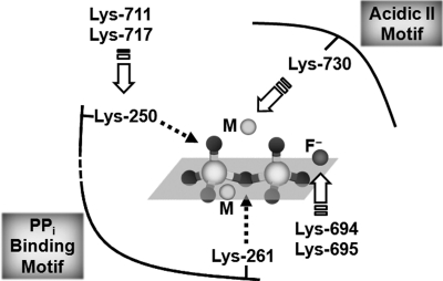
H+-translocating pyrophosphatase (H+-PPase; EC 3.6.1.1) drives proton transport against an electrochemical potential gradient by hydrolyzing pyrophosphate (PPi) and is found in various endomembranes of higher plants, bacteria, and some protists. H+-PPase contains seven highly conserved lysines. We examined the functional roles of these lysines, which are, for the most part, found in the cytosolic regions of mung bean H+-PPase by site-directed mutagenesis. Construction of mutants that each had a cytosolic and highly conserved lysine substituted with an alanine resulted in dramatic drops in the PPi hydrolytic activity. The effects caused by ions on the activities of WT and mutant H+-PPases suggest that Lys-730 may be in close proximity to the Mg2+-binding site, and the great resistance of the K694A and K695A mutants to fluoride inhibition suggests that these lysines are present in the active site. The modifier fluorescein 5'-isothiocyanate (FITC) labeled a lysine at the H+-PPase active site but did not inhibit the hydrolytic activities of K250A, K250N, K250T, and K250S, which suggested that Lys-250 is essential for substrate binding and may be involved in proton translocation. Analysis of tryptic digests indicated that Lys-711 and Lys-717 help maintain the conformation of the active site. Proteolytic evidence also demonstrated that Lys-250 is the primary target of trypsin and confirmed its crucial role in H+-PPase hydrolysis.


Figures

FIGURE 1.
FIGURE 1.
Topology and sequence alignment results for the cytosolic lysines of mung bean H+-PPase. The topology was predicted on the basis of the Streptomyces coelicolor H+-PPase topology (9) with minor modifications. Each cytosolic lysine, with its position number given, is marked with a black circle, highly conserved; a gray circle, moderately conserved; white circle, less or not conserved. Sequence alignment was obtained by ClustalW program (14). Sequences of the H+-PPases from eukaryotes are as follows: Arabidopsis thaliana (NCB accession number P31414), Chara corallina (NCB accession number BAA36841), Oryza sativa (NCB accession number AAQ19328), Triticum aestivum (NCB accession number ABX10014), Vigna radiata (NCB accession number P21616), and Vitis vinifera (NCB accession number CAD89675). Sequences of the H+-PPases from bacteria are as follows: Rhodospirillum rubrum (NCB accession number O68460) and S. coelicolor (NCB accession number Q9X91). Sequences of the H+-PPases from archaea are as follows: Methanococcoides burtonii (NCB accession number YP_565684), Methanosarcina acetivorans (NCB accession number NP_618751), Methanosarcina mazei (NCB accession number NP_632725), and Pyrobaculum aerophilum (NCB accession number AAF01029).
FIGURE 2.
FIGURE 2.
Heterologous expression of mung bean H+-PPase in yeast. A, WT mung bean H+-PPase expression in S. cerevisiae. Microsomal preparations (1st and 2nd lanes) and purified mung bean H+-PPase (3rd lane) (35) were subjected to SDS-PAGE and silver stain (bottom panel) and then immunoblotted with polyclonal anti-H+-PPase antibody. Top panel, lane 1, microsomal fraction of S. cerevisiae that contained a virgin vector; lane 2, microsomal fraction of S. cerevisiae that contained the WT mung bean H+-PPase expression vector; lane 3, purified WT mung bean H+-PPase (35). B, H+ translocating activity of heterologously expressed H+-PPase. The membrane fractions from S. cerevisiae transformed with a WT or virgin vector were assayed for PPi-dependent H+ translocation activity as described under “Experimental Procedures.” C, activity assay of heterologously expressed H+-PPase. The membrane fractions from S. cerevisiae transformed with a WT or virgin vector were assayed for PPi hydrolytic activity as described under “Experimental Procedures.” The concentrations used were 50 mm K+, 100 mm Na+, 100 μm Ca2+, and 5 mm F. Each value is the mean ± S.D. of at least three independent experiments.
FIGURE 3.
FIGURE 3.
Expression and enzymatic activities of the mung bean H+-PPase Lys → Ala mutants. Microsomes were prepared from S. cerevisiae BJ2168 cells expressing WT or a mutated H+-PPase as indicated. A, Western blot. B, PPi hydrolysis, PPi-dependent H+ translocation, and the coupling ratio (top to bottom). The values of the enzymatic activity (29.9 ± 3.8 μmol of PPi hydrolyzed per mg of protein· h), proton translocation (32.7 ± 0.8 × 10³ ΔF%/mg of protein·min), and the coupling ratio for WT were set to 100%. The coupling ratio is the ratio of the initial proton-pumping rate to that of PPi hydrolysis, (ΔF%/min)/(μmol of PPi hydrolyzed per min). Each relative value is the mean ± S.D. of at least three independent experiments. The mutants are classified into three subgroups: I, highly conserved; II, moderately conserved; III, less or not conserved. **, not detectable.
FIGURE 4.
FIGURE 4.
FITC inhibition of Lys → Ala H+-PPase mutants. Microsomes (75 μg) containing WT or a mutant H+-PPase were incubated with 10 mm FITC at 37 °C for 10 min. Each reaction was stopped by diluting the reaction mixture 20-fold with 30 mm MOPS-KOH (pH 7.9), 1 mm Na4PPi, 1 mm MgSO4, 50 mm KCl, 0.5 mm NaF, 1.5 μg/ml gramicidin D. Activities were determined after 10 min of incubation. The remaining activity (%) is reported as the ratio of the enzymatic activities for a protein after it had or had not been subjected to FITC treatment. The PPi hydrolytic activity of the WT control was ∼109.9 ± 0.8 μmol of PPi hydrolyzed per mg protein·h. **, not detectable.
FIGURE 5.
FIGURE 5.
Properties of the Lys-250 H+-PPase mutants. A, expression levels and PPi hydrolytic activities. The expression levels and specific activities of Lys-250 mutants were examined by Western blotting and activity assays, respectively. The residues selected to substitute for Lys-250 were determined by their high score in Dayhoff matrices (36). B, PPi-dependent H+ translocation. C, coupling ratios. The H+ translocation and coupling ratios were determined as described under “Experimental Procedures.” The enzymatic activity (32.9 ± 1.5 μmol of PPi hydrolyzed per mg of protein·h), proton translocation (28.8 ± 0.5 × 10³ (ΔF%/mg of protein·min)), and coupling ratio of WT were each set to 100%. The coupling ratio is the ratio of the initial proton-pumping rate to that of PPi hydrolysis (ΔF%/min)/(μmol of PPi hydrolyzed per min). **, not detectable.
FIGURE 6.
FIGURE 6.
FITC inhibition and tryptic digestion of Lys-250 H+-PPase mutants. A, FITC inhibition of the Lys-250 mutants. Microsomes each enriched in one of the H+-PPase mutants were or were not treated with 10 mm FITC. The remaining activity (%) is reported as the ratio of the activities for a protein after it had or had not been subjected to FITC treatment. The PPi hydrolytic activity of the WT control was 99.4 ± 3.1 μmol of PPi hydrolyzed per mg of protein·h. B, proteolysis assay of K250R. The tryptic digests of WT and K250R were characterized by Western blotting under the conditions shown in the upper panel. Membrane fractions (30 μg) containing H+-PPase were incubated with l-1-tosylamido-2-phenylethyl chloromethyl ketone-treated trypsin in the presence and absence of 2 mm Mg-PPi as described under “Experimental Procedures.”
FIGURE 7.
FIGURE 7.
Trypsin digestion of Lys → Ala H+-PPase mutants. Membrane fractions (30 μg) containing WT or a mutant H+-PPase were incubated with l-1-tosylamido-2-phenylethyl chloromethyl ketone-treated trypsin in the presence and absence of 2 mm Mg-PPi as described under “Experimental Procedures.” H+-PPases that were not digested by trypsin were visualized by immunoblotting. A, group I mutants; B, group II mutants. The control (C) is WT that was not treated with trypsin. V is microsomal fraction of S. cerevisiae that contained a virgin vector.
FIGURE 8.
FIGURE 8.
Proposed model for essential lysines in substrate binding of H+-PPase. The schematic shows the functional lysines involved in substrate binding based on the findings in this work and the crystal structure of soluble PPase (see Ref. or see Protein Data Bank code 2AUU). The lysines are presented with their position numbers in mung bean H+-PPase. Dotted arrowheads indicate the direct interaction caused by Lys-250 and Lys-261, and white arrowheads show the indirect effects exerted on substrate complex by other lysines indicated. The substrate complex is presented as stick-and-ball, where M and F indicate Mg2+ and fluoride, respectively.

Similar articles

Cited by

References

    1. Maeshima M. (2000) Biochim. Biophys. Acta 1465, 37–51 - PubMed
    1. Rea P. A., Poole R. J. (1993) Annu. Rev. Plant Physiol. Plant Mol. Biol. 44, 157–180
    1. Li J., Yang H., Peer W. A., Richter G., Blakeslee J., Bandyopadhyay A., Titapiwantakun B., Undurraga S., Khodakovskaya M., Richards E. L., Krizek B., Murphy A. S., Gilroy S., Gaxiola R. A. (2005) Science 310, 121–125 - PubMed
    1. Guo S., Yin H., Zhang X., Zhao F., Li P., Chen S., Zhao Y., Zhang H. (2006) Plant Mol. Biol. 60, 41–50 - PubMed
    1. Park S., Li J., Pittman J. K., Berkowitz G. A., Yang H., Undurraga S., Morris J., Hirschi K. D., Gaxiola R. A. (2005) Proc. Natl. Acad. Sci. U.S.A. 102, 18830–18835 - PMC - PubMed

Publication types

MeSH terms