. 2011 Jul 1;91(1):27-36.
doi: 10.1093/cvr/cvr042. Epub 2011 Feb 3.

Involvement of vascular peroxidase 1 in angiotensin II-induced vascular smooth muscle cell proliferation

Affiliations

Involvement of vascular peroxidase 1 in angiotensin II-induced vascular smooth muscle cell proliferation

Ruizheng Shi et al. Cardiovasc Res. .

Abstract

Aims: Vascular peroxidase 1 (VPO1) is a newly identified haem-containing peroxidase that catalyses the oxidation of a variety of substrates by hydrogen peroxide (H(2)O(2)). Considering the well-defined effects of H(2)O(2) on the vascular remodelling during hypertension, and that VPO1 can utilize H(2)O(2) generated from co-expressed NADPH oxidases to catalyse peroxidative reactions, the aims of this study were to determine the potential role of VPO1 in vascular remodelling during hypertension.

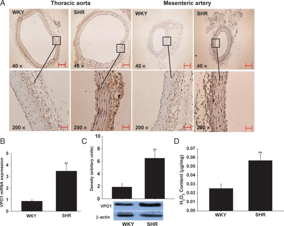
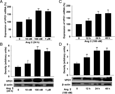
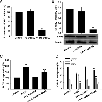
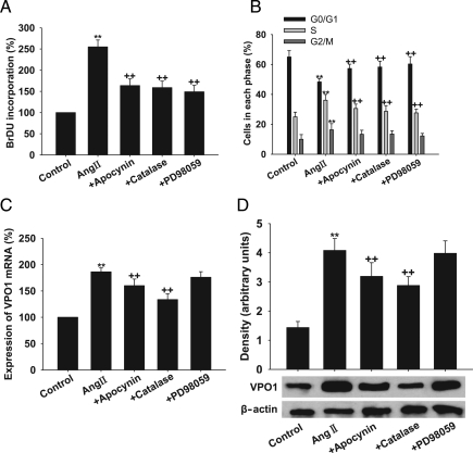
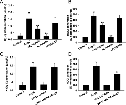
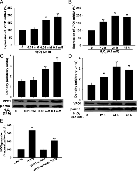
Methods and results: The vascular morphology and the expression of VPO1 in arterial tissues of spontaneously hypertensive rats and Wistar-Kyoto rats were assessed. The VPO1 expression was significantly increased concomitantly with definite vascular remodelling assessed by evaluating the media thickness, lumen diameter, media thickness-to-lumen diameter ratio and mean nuclear area in artery media in spontaneously hypertensive rats. In addition, in cultured rat aortic smooth muscle cells we found that the angiotensin II-mediated cell proliferation was inhibited by knockdown of VPO1 using small hairpin RNA. Moreover, the NADPH oxidase inhibitor, apocynin, and the hydrogen peroxide scavenger, catalase, but not the ERK1/2 inhibitor, PD98059, attenuated angiotensin II-mediated up-regulation of VPO1 and generation of hypochlorous acid.

Conclusion: VPO1 is a novel regulator of vascular smooth muscle cell proliferation via NADPH oxidase-H(2)O(2)-VPO1-hypochlorous acid-ERK1/2 pathways, which may contribute to vascular remodelling in hypertension.


Figures

Figure 1
Figure 1
Expression of VPO1 and content of H2O2 in arterial tissues in rats. (A) The immunohistochemistry analysis of VPO1 expression in thoracic aorta and mesenteric artery from SHRs and WKY rats. Scale bar represents 50 µm. (B) The mRNA expression of VPO1 in thoracic aorta by real-time PCR analysis. (C) The protein expression of VPO1 in thoracic aorta by western blot analysis. (D) The H2O2 content in thoracic aorta. SHRs, spontaneously hypertensive rats; WKY, Wistar–Kyoto rats. n = 10; **P< 0.01 vs. SHR.
Figure 2
Figure 2
Expression of VPO1 in response to Ang II stimulation in A10 VSMCs and the effect of knockdown of VPO1 by shRNA. (AD) Ang II up-regulated the expression of VPO1 (mRNA and protein levels analysed by real-time PCR and western blotting, respectively) in a time- and concentration-dependent manner. **P < 0.01 vs. Ang II 0 nM or 0 h. n = 3. Data are representative of three independent experiments.
Figure 3
Figure 3
Effect of VPO1 knockdown on Ang II-induced proliferation of A10 VSMCs. (A and B) VPO1-shRNA successfully decreased VPO1 mRNA and protein expression, respectively. **P < 0.01 vs. control (C-shRNA). (C) BrDU incorporation. (D) Cell cycle analysis by flow cytometry. Control, wild-type cells; Ang II, wild-type cells treated with 100 nmol/L Ang II for 24 h; VPO1-shRNA, cells transfected with VPO1-shRNA; VPO1-shRNA + Ang II, VPO1-shRNA-tranfected cells treated with 100 nmol/L Ang II for 24 h. **P < 0.01 vs. control; ++P< 0.01 vs. Ang II. n = 3. Data are representative of three independent experiments.
Figure 4
Figure 4
Effect of apocynin, catalase, and PD98059 on VPO1 expression and cell proliferation in Ang II-treated A10 VSMCs. (A) BrdU incorporation. (B) Cell cycle analysis by flow cytometry. (C) VPO1 mRNA measured by real-time PCR analysis. (D) VPO1 expression measured by western blot analysis. Control, wild-type cells; Ang II, wild-type cells treated with 100 nmol/L Ang II for 24 h; +apocynin, cells pre-treated with apocynin (400 µmol/L, 20 min) before 100 nmol/L Ang II for 24 h; +catalase, cells pre-treated with catalase (300 nmol/L, 2 h) before 100 nmol/L Ang II for 24 h; +PD98059, cells pre-treated with PD98059 (10 µmol/L) for 30 min before 100 nmol/L Ang II for 24 h. **P < 0.01 vs. control, ++P< 0.01 vs. Ang II. n = 3. Data are representative of three independent experiments.
Figure 5
Figure 5
Effect of apocynin, catalase, PD98059, and VPO1-shRNA on H2O2 and HOCl levels in Ang II-treated A10 VSMCs. (A) Apocynin, catalase, and PD98059 attenuated H2O2 levels. (B) Apocynin, catalase, and PD98059 attenuated the HOCl levels. (C) VPO1-shRNA attenuated H2O2 levels. (D) VPO1-shRNA attenuated HOCl levels. Control, wild-type cells; Ang II, wild-type cells treated with 100 nmol/L Ang II for 24 h; +apocynin, cells pre-treated with apocynin (400 µmol/L, 20 min) before 100 nmol/L Ang II for 24 h; +catalase, cells pre-treated with catalase (300 nmol/L, 2 h) before 100 nmol/L Ang II for 24 h; +PD98059, cells treated with PD98059 (10 µmol/L) for 30 min before 100 nmol/L Ang II for 24 h; VPO1-shRNA, cells transfected with VPO1-shRNA; VPO1-shRNA + Ang II: VPO1-shRNA transfected cells treated with 100 nmol/L Ang II for 24 h. **P < 0.01 vs. control; ++P< 0.01 vs. Ang II. n = 3. Data are representative of three independent experiments.
Figure 6
Figure 6
VPO1 expression and HOCl levels in response to H2O2 in A10 VSMCs. (AD) H2O2 up-regulated the expression of VPO1 (mRNA and protein analysed by real-time PCR and western blot, respectively) in a time- and concentration-dependent manner. **P < 0.01 vs. H2O2 0 mM or 0 h. (E) VPO1-shRNA successfully decreased HOCl generation. **P < 0.01 vs. control, ++P< 0.01 vs. H2O2. n = 3. Data are representative of three independent experiments.

Comment in

Similar articles

Cited by

References

    1. Gibbons GH, Dzau VJ. The emerging concept of vascular remodeling. N Engl J Med. 1994;330:1431–1438. doi:10.1056/NEJM199405193302008. - DOI - PubMed
    1. Dzau VJ, Gibbons GH, Morishita R, Pratt RE. New perspectives in hypertention research: potentials of vascular biology. Hypertension. 1994;23:1132–1140. - PubMed
    1. Zafari AM, Ushio-Fukai M, Akers M, Yin Q, Shah A, Harrison DG, et al. Role of NADH/NADPH oxidase-derived H2O2 in angiotensin II-induced vascular hypertrophy. Hypertension. 1998;32:488–495. - PubMed
    1. Zhang M, Dong Y, Xu J, Xie Z, Wu Y, Song P, et al. Thromboxane receptor activates the AMP-activated protein kinase in vascular smooth muscle cells via hydrogen peroxide. Circ Res. 2008;102:328–337. doi:10.1161/CIRCRESAHA.107.163253. - DOI - PMC - PubMed
    1. Blanc A, Pandey NR, Srivastava AK. Synchronous activation of ERK 1/2, p38mapk and PKB/Akt signaling by H2O2 in vascular smooth muscle cells: potential involvement in vascular disease. Int J Mol Med. 2003;11:229–234. - PubMed

Publication types

MeSH terms