

Oxygen-dependent quenching of phosphorescence used to characterize improved myocardial oxygenation resulting from vasculogenic cytokine therapy
- PMID: 21292844
- PMCID: PMC3098666
- DOI: 10.1152/japplphysiol.01138.2010
Oxygen-dependent quenching of phosphorescence used to characterize improved myocardial oxygenation resulting from vasculogenic cytokine therapy
Abstract
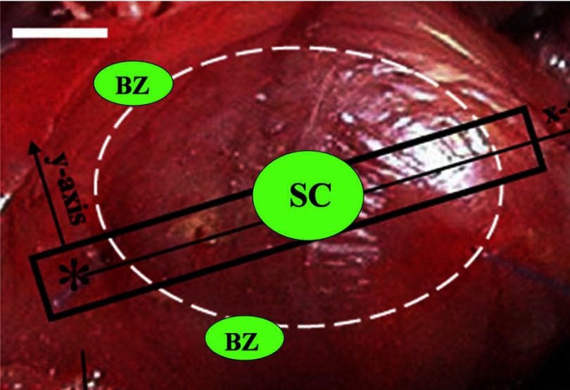
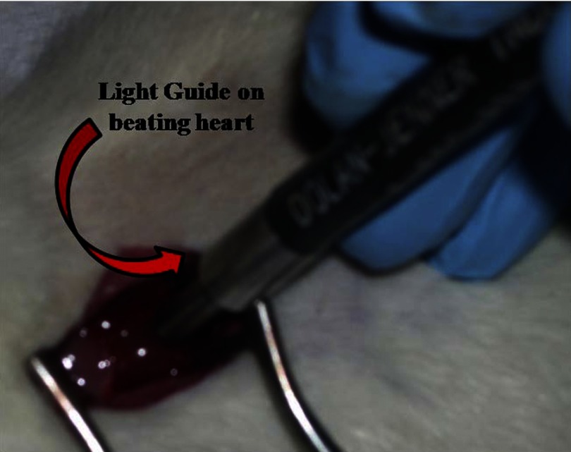
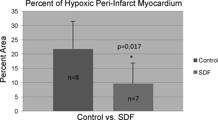
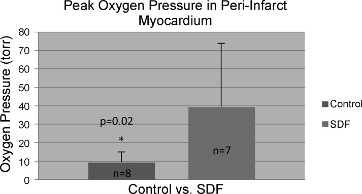
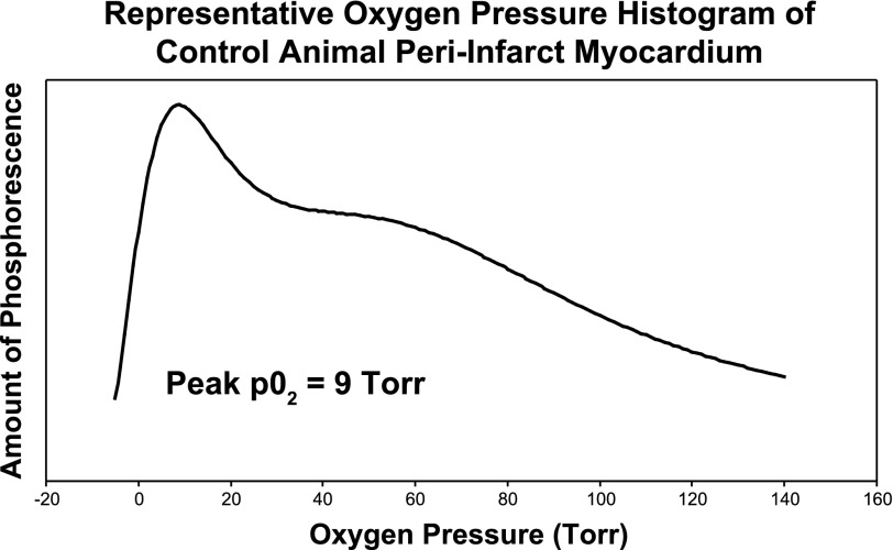
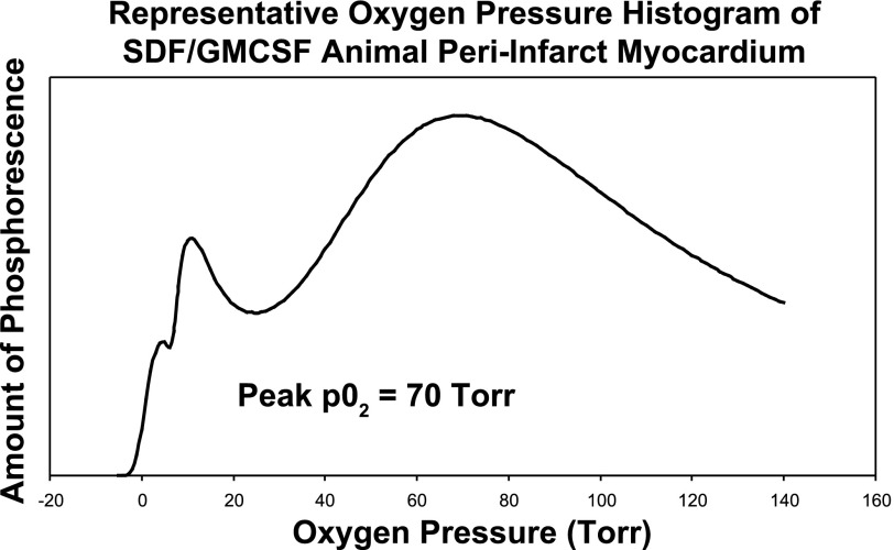
This study evaluates a therapy for infarct modulation and acute myocardial rescue and utilizes a novel technique to measure local myocardial oxygenation in vivo. Bone marrow-derived endothelial progenitor cells (EPCs) were targeted to the heart with peri-infarct intramyocardial injection of the potent EPC chemokine stromal cell-derived factor 1α (SDF). Myocardial oxygen pressure was assessed using a noninvasive, real-time optical technique for measuring oxygen pressures within microvasculature based on the oxygen-dependent quenching of the phosphorescence of Oxyphor G3. Myocardial infarction was induced in male Wistar rats (n = 15) through left anterior descending coronary artery ligation. At the time of infarction, animals were randomized into two groups: saline control (n = 8) and treatment with SDF (n = 7). After 48 h, the animals underwent repeat thoracotomy and 20 μl of the phosphor Oxyphor G3 was injected into three areas (peri-infarct myocardium, myocardial scar, and remote left hindlimb muscle). Measurements of the oxygen distribution within the tissue were then made in vivo by applying the end of a light guide to the beating heart. Compared with controls, animals in the SDF group exhibited a significantly decreased percentage of hypoxic (defined as oxygen pressure ≤ 15.0 Torr) peri-infarct myocardium (9.7 ± 6.7% vs. 21.8 ± 11.9%, P = 0.017). The peak oxygen pressures in the peri-infarct region of the animals in the SDF group were significantly higher than the saline controls (39.5 ± 36.7 vs. 9.2 ± 8.6 Torr, P = 0.02). This strategy for targeting EPCs to vulnerable peri-infarct myocardium via the potent chemokine SDF-1α significantly decreased the degree of hypoxia in peri-infarct myocardium as measured in vivo by phosphorescence quenching. This effect could potentially mitigate the vicious cycle of myocyte death, myocardial fibrosis, progressive ventricular dilatation, and eventual heart failure seen after acute myocardial infarction.
Figures
References
-
- Assmus B, Schachinger V, Teupe C, Britten M, Lehmann R, Dobert N, Grunwald F, Aicher A, Urbich C, Martin H, Hoelzer D, Dimmeler S, Zeiher AM. Transplantation of progenitor cells and regeneration enhancement in acute myocardial infarction (TOPCARE-AMI). Circulation 106: 3009–3017, 2002. - PubMed
-
- Atluri P, Liao GP, Panlilio CM, Hsu VM, Leskowitz MJ, Morine KJ, Cohen JE, Berry MF, Suarez EE, Murphy DA, Lee WMF, Gardner TJ, Sweeney HL, Woo YJ. Neovasculogenic therapy to augment perfusion and preserve viability in ischemic cardiomyopathy. Ann Thorac Surg 81: 1728–1736, 2006. - PubMed
-
- Balcells E, Powers ER, Lepper W, Belcik T, Wei K, Ragosta M, Samady H, Lindner JR. Detection of myocardial viability by contrast echocardiography in acute infarction predicts recovery of resting function and contractile reserve. J Am Coll Cardiol 41: 827–833, 2003. - PubMed
-
- Behnke BJ, Kindig CA, Musch TI, Koga S, Poole DC. Dynamics of microvascular oxygen pressure across the rest-exercise transition in rat skeletal muscle. Respir Physiol 126: 53–63, 2001. - PubMed
Publication types
MeSH terms
Substances
Grants and funding
LinkOut - more resources
Full Text Sources
Medical