

A focus of dogs and Rickettsia massiliae-infected Rhipicephalus sanguineus in California
- PMID: 21292893
- PMCID: PMC3029176
- DOI: 10.4269/ajtmh.2011.10-0355
A focus of dogs and Rickettsia massiliae-infected Rhipicephalus sanguineus in California
Abstract
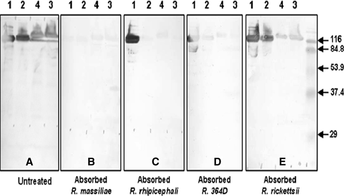
A recurrent focus of Rhipicephalus sanguineus infestation was investigated in a suburban area of southern California after reports of suspected Rocky Mountain spotted fever in two dogs on the same property. Abundant quantities of Rh. sanguineus were collected on the property and repeatedly from each dog, and Rickettsia massiliae DNA was detected by polymerase chain reaction (PCR). Whole blood and serum samples from four dogs were tested by using PCR and microimmunofluorescent assay for antibodies against spotted fever group rickettsiae. Serum samples from all four dogs contained antibodies reactive with R. massiliae, R. rhipicephali, R. rickettsii, and 364D Rickettsia but no rickettsial DNA was detected by PCR of blood samples. Serum cross-absorption and Western blot assays implicated R. massiliae as the most likely spotted fever group rickettsiae responsible for seropositivity. To our knowledge, this is the first detection of R. massiliae in ticks in California.
Figures
References
-
- Beati L, Finidori JP, Gilot B, Raoult D. Comparison of serologic typing, sodium dodecyl sulfate-polyacrylamide gel electrophoresis protein analysis, and genetic restriction fragment length polymorphism analysis for identification of rickettsiae: characterization of two new rickettsial strains. J Clin Microbiol. 1992;30:1922–1930. - PMC - PubMed
-
- Beati L, Raoult D. Rickettsia massiliae sp. nov., a new spotted fever group Rickettsia. Int J Syst Bacteriol. 1993;43:839–840. - PubMed
-
- Babalis T, Tselentis Y, Roux V, Psaroulaki A, Raoult D. Isolation and identification of a rickettsial strain related to Rickettsia massiliae in Greek ticks. Am J Trop Med Hyg. 1994;50:365–372. - PubMed
MeSH terms
LinkOut - more resources
Full Text Sources
