. 2011 Mar 4;331(6021):1188-91.
doi: 10.1126/science.1201476. Epub 2011 Feb 3.

A sorting platform determines the order of protein secretion in bacterial type III systems

Affiliations

A sorting platform determines the order of protein secretion in bacterial type III systems

María Lara-Tejero et al. Science. .

Abstract

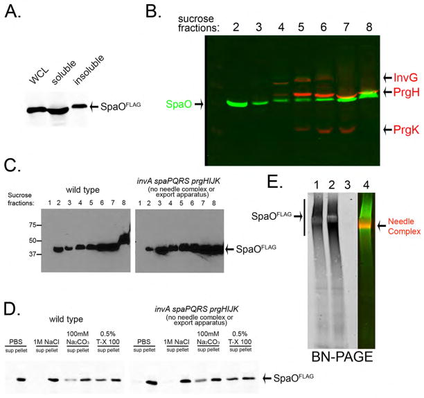
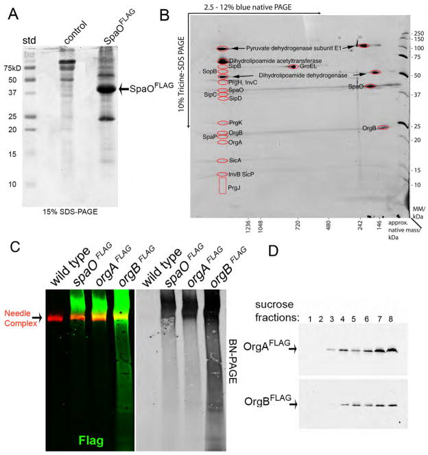
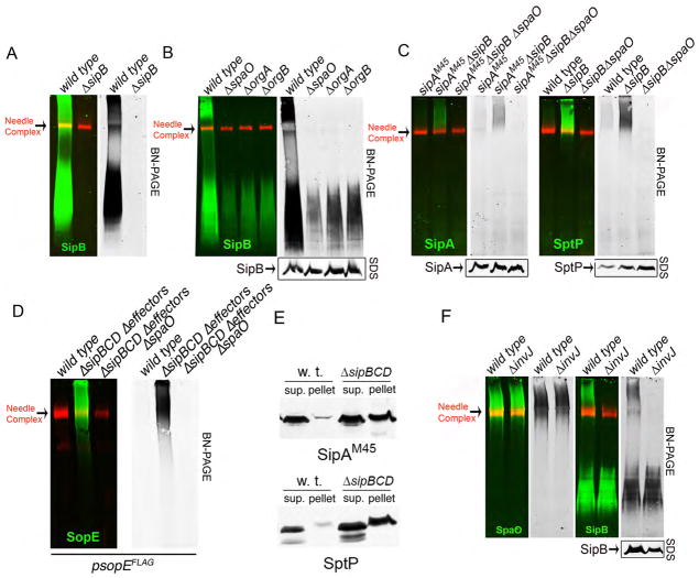
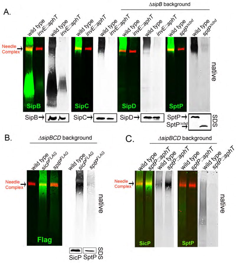
Bacterial type III protein secretion systems deliver effector proteins into eukaryotic cells in order to modulate cellular processes. Central to the function of these protein-delivery machines is their ability to recognize and secrete substrates in a defined order. Here, we describe a mechanism by which a type III secretion system from the bacterial enteropathogen Salmonella enterica serovar Typhimurium can sort its substrates before secretion. This mechanism involves a cytoplasmic sorting platform that is sequentially loaded with the appropriate secreted proteins. The sequential loading of this platform, facilitated by customized chaperones, ensures the hierarchy in type III protein secretion. Given the presence of these machines in many important pathogens, these findings can serve as the bases for the development of novel antimicrobial strategies.


Figures

Fig. 1
Fig. 1. SpaO forms a large molecular weight complex
(A, B) Subcellular fractionation of SpaO (A) A whole cell lysate (WCL) of a S. Typhimurium strain expressing FLAG-epitope-tagged SpaO was separated into soluble and pelletable fractions by high-speed centrifugation and the presence of SpaO in the different fractions was probed by immuno blotting with an anti FLAG antibody. (B) The pellet fraction was subsequently separated by sucrose gradient centrifugation and the different gradient fractions were probed for the presence of SpaO (green) and needle complex components (red) by immuno blotting and imaging with the Odyssey system (Li-Cor Bioscience). (C) Comparison of the distribution of SpaO in wild type and the ΔinvA ΔspaPQRS ΔprgHIJK isogenic mutant derivative, which lacks the needle complex and membrane protein components of the export apparatus. The distribution of SpaO in the different fractions of a sucrose gradient was probed as indicated above. (D) Localization of SpaO after different treatments. Pellet fractions of SpaO obtained from the indicated strains were subjected to different treatments (as indicated) and its localization of high-speed centrifugation was determined by immuno blot analysis of the pellet and soluble fractions. (E) Analysis of the SpaO complex by BN-PAGE. Pellet fractions from a wild-type S. Typhimurium encoding a FLAG-epitope-tagged SpaO (1), a ΔinvA ΔspaPQRS ΔprgHIJK mutant derivative (2), or the untagged wild-type strain (3) were separated by BN-PAGE and analyzed by immuno blot for the presence of SpaO or the needle complex. The simultaneous detection of the SpaO (green) and needle complexes (red) from the sample shown on lane 1 is shown in lane 4. Only the SpaO channel is shown in lanes 1, 2, and 3.
Figure 2
Figure 2. Characterization of the SpaO complex
(A) and (B) Cell lysates of wild-type S. Typhimurium or an isogenic strain expressing FLAG-epitope tagged SpaO were immunoprecipitated with an anti FLAG antibody, separated by SDS-PAGE (A) and interacting proteins were identified by LC-MS/MS (see Fig. S1). Alternatively, the pelletable fraction of a cell lysates from a S. Typhimurium expressing FLAG-epitope tagged SpaO was separated on a sucrose density gradient, SpaO-containing fractions were pooled and SpaO-interacting proteins were immunoprecipitated with an anti-FLAG antibody, separated by 2D-BN-PAGE (B), and the identity of the proteins in the indicated spots (red circles) was established by LC-MS/MS (see Fig. S2). (C) Whole cell lysate of a S. Typhimurium strain expressing functional FLAG-epitope tagged SpaO, OrgA, or OrgB were separated into soluble and pelletable fractions by high-speed centrifugation. Pellet fractions were subsequently applied to a sucrose gradient, relevant fractions pulled, separated by BN-PAGE, and analyzed by immuno blot for the presence of SpaO, OrgA, or OrgB (green) or the needle complex (red). The simultaneous detection of the needle complex and SpaO, OrgA, or OrgB is shown in the color panel while only the detection of SpaO, OrgA, or OrgB is shown on the right (black and white) panel. (D) Subcellular fractionation of OrgA and OrgB. A whole cell lysate of a S. Typhimurium strain expressing FLAG-epitope-tagged OrgA or OrgB were separated into soluble and pelletable fractions by high-speed centrifugation. The pellet fractions were subsequently separated by sucrose gradient centrifugation and the different gradient fractions were probed for the presence of OrgA or OrgB by immuno blotting.
Figure 3
Figure 3. The SpaO/OrgA/OrgB platform can be alternatively loaded with different substrates of the type III secretion system
(A and B) Whole cell lysates of wild-type S. Typhimurium or the ΔsipB, ΔspaO, ΔorgA or ΔorgB mutants were separated into soluble and pelletable fractions by high-speed centrifugation. Pellets were further fractionated by sucrose gradient centrifugation, relevant fraction pooled, separated by BN-PAGE, and analyzed by immuno blot for the presence of SipB (green) or the needle complex (red). Left panels show the simultaneous detection of SipB and the needle complex and right (black and white) panels show only the detection of SipB. The lower panel shows SDS-PAGE immuno-blot analysis of SipB in equal amounts of whole cell lysates of the indicated strains. (C) (D) and (E) Samples from the indicated strains were separated into soluble (sup) and pelletable (pellet) fractions by high-speed centrifugation and separated by SDS-PAGE (E). Alternatively, the pellet fraction was further separated on a sucrose gradient, and the relevant fractions pooled and separated by BN-PAGE (C) and (D). Left panels in (C) and (D) show the simultaneous detection of SipA, SptP, (C), or SopE (D) (green) and the needle complexes (red) and right (black and white) panels show only the channel corresponding to the detection of the respective effector proteins. Lower panels show SDS-PAGE immuno-blot analysis of the indicated proteins in equal amounts of whole cell lysates of the indicated strains (C) and (D). (F) Samples from wild-type S. Typhimurium or a Δ invJ mutant expressing a FLAG-tagged SpaO were prepared as indicated above and separated by BN-PAGE. The left panels show the simultaneous detection of SpaO or SipB (green) and the needle complexes (red) and the right (black and white) panels shows only the channel corresponding to the detection of SpaO or SipB. The lower panel shows SDS-PAGE immuno-blot analysis of SipB in equal amounts of whole cell lysates of the indicated strains.
Fig. 4
Fig. 4. The type III secretion chaperones are required for the loading of translocases and effector protein onto the SpaO/OrgA/OrgB platform
(A) Samples from the wild-type S. Typhimurium, a mutant lacking the translocase-chaperone InvE, or a strain expressing a mutant of SptP lacking its chaperone-binding domain (SptPΔ35–161;indicated as SptPΔcbd) were prepared as indicated above and the presence of the translocases SipB, SipC and SipD and the effector protein SptP in the SpaO/OrgA/OrgB complex was examined by BN-PAGE and immuno blot analysis. A S. Typhimurium lacking SipB was used as a background strain for these experiments to enhance the detection of SipD or SptP. In all cases the left panel show the simultaneous detection of the indicated effector or translocase (green) and the needle complexes (red) and right (black and white) panels show only the channel corresponding to the detection of the respective effector or translocase proteins. Lower panels show SDS-PAGE immuno-blot analysis of the indicated proteins in equal amounts of whole cell lysates of the indicated strains. (B) Samples from wild-type S. Typhimurium, a strain expressing FLAG epitope tagged SicP, the chaperone for SptP, or a strain expressing FLAG-tagged SptP, were prepared as indicated above and separated by BN-PAGE. The left panel shows the simultaneous detection of SicPFLAG or SptPFLAG (green) and the needle complexes (red) and right (black and white) panel shows only the channel corresponding to the detection of SicPFLAG or SptPFLAG. The lower panels show SDS-PAGE immuno-blot analysis of the indicated proteins in equal amounts of whole cell lysates of the indicated strains. (C) Samples from the indicated strains were prepared as described above and separated by BN-PAGE. The left panel shows the simultaneous detection of SicPFLAG or SptP (green) and the needle complexes (red) and right (black and white) panels show only the channel corresponding to the detection of SicPFLAGor SptP.

Comment in

References

    1. Galan JE, Cossart P. Curr Opin Microbiol. 2005;8:1. - PubMed
    1. Galan JE, Wolf-Watz H. Nature. 2006;444:567. - PubMed
    1. Alvarez-Martinez C, Christie P. Microbiol Mol Biol Rev. 2009;73:775. - PMC - PubMed
    1. Pukatzki S, McAuley S, Miyata S. Curr Opin Microbiol. 2009;12:11. - PubMed
    1. Cornelis G. Nat Rev Microbiol. 2006;4:811. - PubMed

Publication types

MeSH terms

Substances