. 2011 Feb 4;331(6017):571-5.
doi: 10.1126/science.1198461.

Protein native-state stabilization by placing aromatic side chains in N-glycosylated reverse turns

Affiliations

Protein native-state stabilization by placing aromatic side chains in N-glycosylated reverse turns

Elizabeth K Culyba et al. Science. .

Erratum in

  • Science. 2012 Mar 2;335(6072):1042

Abstract

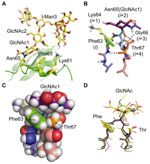
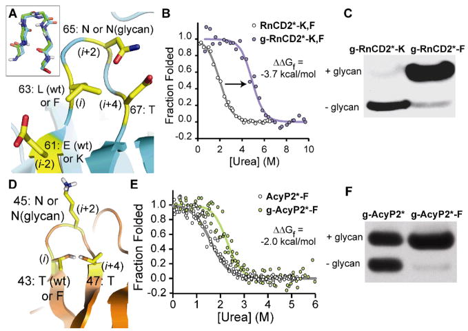
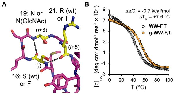
N-glycosylation of eukaryotic proteins helps them fold and traverse the cellular secretory pathway and can increase their stability, although the molecular basis for stabilization is poorly understood. Glycosylation of proteins at naïve sites (ones that normally are not glycosylated) could be useful for therapeutic and research applications but currently results in unpredictable changes to protein stability. We show that placing a phenylalanine residue two or three positions before a glycosylated asparagine in distinct reverse turns facilitates stabilizing interactions between the aromatic side chain and the first N-acetylglucosamine of the glycan. Glycosylating this portable structural module, an enhanced aromatic sequon, in three different proteins stabilizes their native states by -0.7 to -2.0 kilocalories per mole and increases cellular glycosylation efficiency.


Figures

Figure 1
Figure 1
(A) Ribbon diagram of the published NMR structure of HsCD2ad (PDB: 1GYA) (16). The N-glycan, Asn65, Phe63, and Lys61 are highlighted in yellow. (B) Stick and (C) space-filling representations of the type I β-bulge turn in HsCD2ad; the (i+2)-position Asn65(GlcNAc1) packs against the i-position Phe63, and the i+4 Thr67. Dashed lines indicate hydrogen bonds. (D) Glycosylated type I β-bulge turns with an i-position Phe from known proteins in the PDB (green: HsCD2ad; blue: 1G82; orange: 1FJR; red: 1ICF; and black: 3DMK). All structures rendered in Pymol (www.pymol.org).
Figure 2
Figure 2
(A) Ribbon diagram of a type I β-bulge turn in the known crystal structure of RnCD2ad (PDB: 1HNG) (19), rendered in Pymol; inset shows the backbone alignment of this turn with the corresponding reverse turn from HsCD2ad. Overlaid text shows the amino acids to be perturbed at positions 61 (i-2), 63 (i), 65 (i+2), and 67 (i+4), which are highlighted in yellow. wt, wild type (B) Fluorescence-monitored urea denaturation curve for glycoprotein g-RnCD2*-K,F (purple circles) and non-glycosylated RnCD2*-K,F (white circles). Fits of the data (18) appear as purple and gray lines, respectively. (C) Western blot (anti-FLAG tag) of conditioned media from infected Sf9 cells expressing g-RnCD2*-K and g-RnCD2*-F. (D) Ribbon diagram of a reverse turn in the known NMR structure of horse AcyP2 (PDB: 1APS; 95% identical to human, with 100% identity in residues 43 to 47) (22), rendered in Pymol. Overlaid text shows the amino acids to be perturbed at positions 43 (i), 45 (i+2), and 47 (i+4), which are highlighted in yellow. (E) Fluorescence-monitored urea denaturation curve for glycoprotein g-AcyP2*-F (green circles) and non-glycosylated AcyP2*-F (open circles). Fits of the data to a two-state folding model (18) appear as green and gray lines, respectively. (F) Western blot (anti-FLAG tag) of conditioned media from infected Sf9 cells expressing g-AcyP2* and g-AcyP2*-F.
Figure 3
Figure 3
(A) Stick representation of loop 1 in the known crystal structure of Pin WW (PDB: 1PIN) (26), rendered in Pymol. Overlaid text shows the amino acids to be perturbed at positions 16 (i), 19 (i+3), and 21 (i+5), which are highlighted in yellow. Dashed lines indicate hydrogen bonds in wild-type Pin WW. (B) Variable temperature circular dichroism data (monitored at 227 nm) for glycoprotein g-WW-F,T (orange circles) and non-glycosylated WW-F,T (open circles). Fits of the data to a two-state folding model (18) are shown in orange and gray lines, respectively.

References

    1. Kornfeld R, Kornfeld S. Annu Rev Biochem. 1985;54:631. - PubMed
    1. Molinari M. Nat Chem Biol. 2007;3:313. - PubMed
    1. Helenius A, Aebi M. Science. 2001;291:2364. - PubMed
    1. Glozman R, et al. J Cell Biol. 2009;184:847. - PMC - PubMed
    1. Wormald MR, et al. Eur J Biochem. 1991;198:131. - PubMed

Publication types

MeSH terms