. 2011 Feb 22;108(8):3228-33.
doi: 10.1073/pnas.1015953108. Epub 2011 Feb 3.

Ribosome recycling depends on a mechanistic link between the FeS cluster domain and a conformational switch of the twin-ATPase ABCE1

Affiliations

Ribosome recycling depends on a mechanistic link between the FeS cluster domain and a conformational switch of the twin-ATPase ABCE1

Dominik Barthelme et al. Proc Natl Acad Sci U S A. .

Abstract

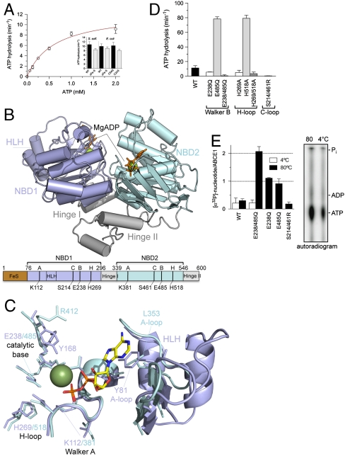
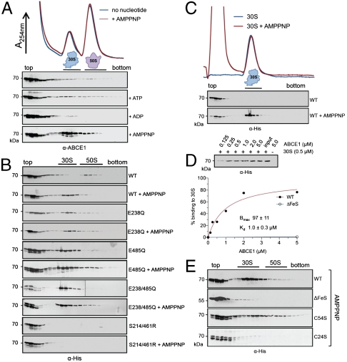
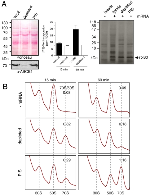
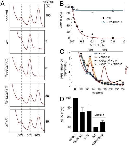
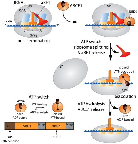
Despite some appealing similarities of protein synthesis across all phyla of life, the final phase of mRNA translation has yet to be captured. Here, we reveal the ancestral role and mechanistic principles of the newly identified twin-ATPase ABCE1 in ribosome recycling. We demonstrate that the unique iron-sulfur cluster domain and an ATP-dependent conformational switch of ABCE1 are essential both for ribosome binding and recycling. By direct (11) interaction, the peptide release factor aRF1 is shown to synergistically promote ABCE1 function in posttermination ribosome recycling. Upon ATP binding, ABCE1 undergoes a conformational switch from an open to a closed ATP-occluded state, which drives ribosome dissociation as well as the disengagement of aRF1. ATP hydrolysis is not required for a single round of ribosome splitting but for ABCE1 release from the 30S subunit to reenter a new cycle. These results provide a mechanistic understanding of final phases in mRNA translation.


Conflict of interest statement

The authors declare no conflict of interest.

Figures

Fig. 1.
Fig. 1.
Structure and function of ABCE1. (A) ATPase activity of ABCE1 (5 μM) at 80 °C. Experiments were performed in triplicates. Data represented as mean ± SEM are fitted to the Michaelis–Menten equation. The ATP turnover rate kcat of various FeS cluster mutants (5 μM) is shown as insert. (B) X-ray structure of ABCE1ΔFeS-E238/485Q from S. solfataricus (side view). In the open ADP-bound state, ABCE1 shows a V-like architecture with NBD1 (pale blue) and NBD2 (cyan) preoriented via the hinge domain (gray). The two bound Mg-ADP molecules are shown as green spheres and orange sticks. Functional domains and critical residues in the Walker A and B motifs, C- and H-loop are illustrated below. (C) Overlay of the conserved motifs and catalytic residues of NBD1 (pale blue) and NBD2 (cyan) with regard to the bound Mg-ADP (ADP as sticks, Mg2+ as green sphere). Notably, the catalytic Glu 238 and 485 are exchanged to Gln in the X-ray structure. (D) ATP turnover rate kcat of ABCE1 mutants (5 μM) with substitutions in conserved motifs (Walker B motifs, H- and C-loop). All experiments were performed in triplicates at 80 °C. (E) Stoichiometry of occluded nucleotides. ABCE1 mutants (5 μM) were incubated with 500 μM ATP (traced with [α-32P]-ATP) at 4 °C (white) or 80 °C (black bars). Free and bound nucleotides were separated by spin-down gel filtration and quantified by Cerenkov counting. Data from two independent experiments performed in triplicates are represented as mean ± SEM. The nucleotides occluded in ABCE1E238/485Q were identified by thin layer chromatography (Right).
Fig. 2.
Fig. 2.
The ATP-occluded state and the FeS cluster domain of ABCE1 are essential for stable ribosome association. (A) Ribosome association of ABCE1 was analyzed by SDG in 100 μL of WCE (15 mg/mL) from S. solfataricus incubated in the presence of different nucleotides at 73 °C for 4 min. Fractions (0.5 mL) were analyzed by SDS-PAGE and immunoblotting using an anti-ABCE1 antibody. (B) Isolated ABCE1 mutants (0.4 μM) were incubated with 100 μL of S. solfataricus WCE (15 mg/mL) with and without 5 mM of AMPPNP and analyzed as described above using an anti-His antibody. (C) Binding of ABCE1WT (1 μM) to isolated 30S particles (1 μM) assayed by SDG analysis in the presence and absence of AMPPNP (5 mM) at 73 °C for 4 min. (D) Ribosome pelleting assays of ABCE1WT and isolated 30S subunits (0.5 μM) reveal a stoichiometric (1∶1) binding in the presence of AMPPNP. The amount of ABCE1WT expected for 100% binding (0.5 μM) is given as input. Data were analyzed by quantitative immunoblotting and fitted according to a one-site binding isotherm. (E) SDG analysis of FeS cluster mutants. Purified ABCE1WT, ABCE1ΔFeS, ABCE1C54S, or ABCE1C24S (0.2 μM of each) was added to 100 μL S. solfataricus WCE (15 mg/mL) and incubated with 5 mM of AMPPNP at 73 °C for 4 min. Fractions were probed with an anti-His antibody.
Fig. 3.
Fig. 3.
ABCE1 acts downstream of translation initiation. (A) Immunodepletion of ABCE1 from S. solfataricus WCE (Left). Immunodepletion by preimmune serum serves as control (PIS). The effect of ABCE1 depletion on protein synthesis was assayed by measuring the total capacity of protein synthesis via [35S]-methionine incorporation in TCA precipitates (Center) and by in vitro translation in lysates programmed for mRNA-104 translation coding for rpl30 (Right). (B) Effect of ABCE1 depletion on 70S formation. After in vitro translation of mRNA-104 for 15 or 60 min at 73 °C, reactions were formaldehyde cross-linked and analyzed by SDG centrifugation. The peak areas (A254 nm) corresponding to 70S and 50S were calculated.
Fig. 4.
Fig. 4.
A conformational switch of ABCE1 powers 70S breakdown. (A) ABCE1 mutants (0.8 μM of each) were added to lysates programmed for translation at 73 °C for 30 min and the amount of 70S particles formed were examined by SDG fractionation. The peak area (A254 nm) of 50S and 70S ribosomes was calculated. The 70S/50S ratio of the control reaction was set to 100%. (B) 70S breakdown at different concentrations of ABCE1. Reactions were performed and analyzed as described in (A). The initial 70S/50S ratio was set to 100%. (C) Preinitiation complex formation of lysates programmed with [32P]-labeled mRNA-104 was visualized by Cerenkov counting of the corresponding SDG fractions (10–20% sucrose). ABCE1WT (0.8 μM) was added to these lysates in combination with different nucleotides (2 mM of each). (D) Recombinant ABCE1, aIF6 or GMPPNP were added to lysates programmed for in vitro translation. After 2-min incubation, samples were cross-linked on ice and analyzed by SDG fractionation. Addition of ABCE1WT but also the ATPase inactive mutant ABCE1E238/485Q leads to a significantly larger 70S breakdown than GMPPNP (2 mM) or aIF6 (5 μM). The initial 70S/50S ratio without additional factors (control) was set to 100%.
Fig. 5.
Fig. 5.
ABCE1 functions synergistically with aRF1 in posttermination ribosome disassembly. (A) Purified ABCE1 and aRF1 (2 μM of each) were incubated in the presence of different nucleotides (5 mM) at 73 °C for 10 min. Coimmunoprecipitation by a polyclonal anti-ABCE1 antibody was performed in Co-IP buffer (in vitro) or in 100 μL of WCE (15 mg/mL). Preimmune serum coupled beads served as a control (mock). The asterisk marks the cross-reaction of the eluted antibody heavy chain. (B) Lysates programmed for translation were pulsed for 2 min with increasing concentrations of ABCE1 alone or in combination with aRF1 (1 μM). After the pulse, samples were cross-linked on ice. Ribosome profiles were analyzed as in Fig. 4B. The initial 70S/50S ratio (no additional ABCE1) was set to 100%. (C) Localization of [32P]-labeled ABCE1WT or ABCE1E238/485Q (0.8 μM of each, left panel) in lysates programmed for translation in the presence of aRF1 (1 μM). The ribosome profile at A254 nm in the presence of ABCE1E238/485Q is shown. After ribosome splitting by ABCE1WT and aRF1 (1 μM of each) in the presence of AMPPNP (Lower), the ribosome association of ABCE1 and aRF1 was analyzed by SDG fractionation and immunoblotting by an anti-His antibody. (D) In vitro 70S ribosome breakdown. Isolated 70S ribosomes from T. celer (1 μM) were incubated with ABCE1 (1 μM) in the presence and absence of aRF1 (1 μM) for 4 min at 73 °C and different nucleotides (2 mM of each). Ribosome reassociation was prevented by addition of aIF6 (5 μM). Data were analyzed by SDG fractionation as described in Fig. 4B. The 70S/50S ratio in the absence of added ABCE1 served as control (100%). Experiments were performed in duplicates.
Fig. 6.
Fig. 6.
Model of ribosome recycling in Archaea. After termination, ABCE1 is recruited to the 70S ribosome by binding to aRF1. ATP and ribosome binding induces a conformational switch into a closed state of ABCE1, which powers ribosome dissociation and release of aRF1. Finally, ATP hydrolysis triggers the dissociation of ABCE1 from the 30S subunit. In addition, the conformational changes that drive 70S ribosome dissociation (ATP-switch) and putative interaction sites with aRF1, 30S and RNA are illustrated.

References

    1. Dever TE. Gene-specific regulation by general translation factors. Cell. 2002;108:545–556. - PubMed
    1. Ramakrishnan V. Ribosome structure and the mechanism of translation. Cell. 2002;108:557–572. - PubMed
    1. Chakraburtty K. Translational regulation by ABC systems. Res Microbiol. 2001;152:391–399. - PubMed
    1. Rees DC, Johnson E, Lewinson O. ABC transporters: The power to change. Nat Rev Mol Cell Biol. 2009;10:218–227. - PMC - PubMed
    1. Schmitt L, Tampé R. Structure and mechanism of ABC transporters. Curr Opin Struct Biol. 2002;12:754–760. - PubMed

Publication types

MeSH terms

Substances

Associated data

LinkOut - more resources