. 2011 Feb 22;108(8):3108-15.
doi: 10.1073/pnas.1019660108. Epub 2011 Feb 3.

Real-time analysis of double-strand DNA break repair by homologous recombination

Affiliations

Real-time analysis of double-strand DNA break repair by homologous recombination

Wade M Hicks et al. Proc Natl Acad Sci U S A. .

Abstract

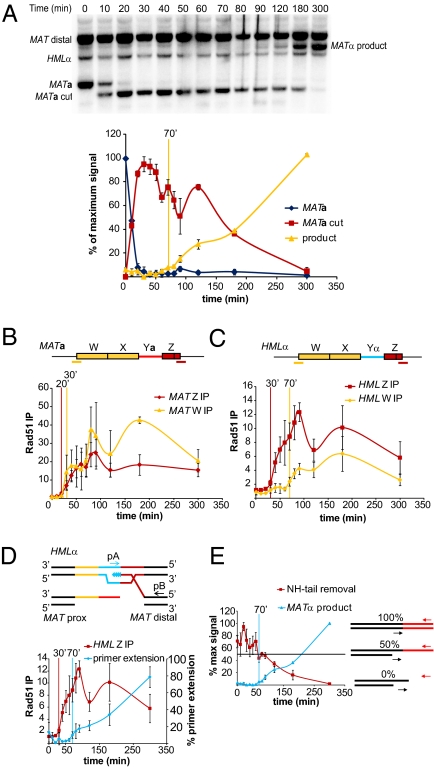
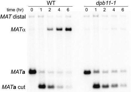
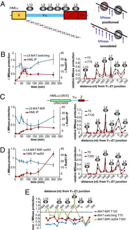
The ability to induce synchronously a single site-specific double-strand break (DSB) in a budding yeast chromosome has made it possible to monitor the kinetics and genetic requirements of many molecular steps during DSB repair. Special attention has been paid to the switching of mating-type genes in Saccharomyces cerevisiae, a process initiated by the HO endonuclease by cleaving the MAT locus. A DSB in MATa is repaired by homologous recombination--specifically, by gene conversion--using a heterochromatic donor, HMLα. Repair results in the replacement of the a-specific sequences (Ya) by Yα and switching from MATa to MATα. We report that MAT switching requires the DNA replication factor Dpb11, although it does not require the Cdc7-Dbf4 kinase or the Mcm and Cdc45 helicase components. Using Southern blot, PCR, and ChIP analysis of samples collected every 10 min, we extend previous studies of this process to identify the times for the loading of Rad51 recombinase protein onto the DSB ends at MAT, the subsequent strand invasion by the Rad51 nucleoprotein filament into the donor sequences, the initiation of new DNA synthesis, and the removal of the nonhomologous Y sequences. In addition we report evidence for the transient displacement of well-positioned nucleosomes in the HML donor locus during strand invasion.


Conflict of interest statement

The authors declare no conflict of interest.

Figures

Fig. 1.
Fig. 1.
Schematic of S. cerevisiae mating type loci on chromosome III and the SDSA model of MAT switching. (A) Chromosome III contains three mating type genes, the expressed MAT locus and HML and HMR, which are heterochromatic and silent. MATa cells switch using HMLα, whereas MATα cells recombine with HMRa. This preference is controlled by the RE element. (B) Individual steps of the DSB repair mechanism are numbered on the left. 1: MAT switching is initiated by an HO-induced DSB at the Ya-Z1 junction within MATa. 2: 5′–3′ resection creates ssDNA with 3′ ends to which Rad51 (yellow circles) is recruited. 3: The Rad51 filament searches for homologous dsDNA. 4: Upon MAT-HML synapsis, new DNA synthesis is primed from the free 3′ end of the invading strand. 5: The nascent strand is dissociated from the donor and anneals with homologous sequences on the left side of the break. 6: The nonhomologous 3′ tail is clipped off, and new DNA synthesis begins at the free 3′ ends to fill in the single-stranded gaps. 7: DSB repair is completed by ligation of the filled-in ends. In the SDSA model, all newly synthesized DNA appears at the repaired locus, whereas the donor locus remains unmodified.
Fig. 2.
Fig. 2.
High-resolution kinetics of MATa switching to MATα. (A) Representative Southern blot showing MAT switching progression. Purified gDNA was digested with StyI. Graph shows quantitative densitometric analysis from two independent experiments; error bars represent ranges. Vertical yellow bar indicates the time point at which product formation becomes detectable over background. (B) Rad51 ChIP analysis at MATa. (C) Rad51 ChIP analysis at HMLα. Red and yellow bars underneath the MATa and HMLα denote positions of primer pairs used to detect Rad51 recruitment to these regions. Rad51 ChIP data were normalized to ARG5,6. Each ChIP experiment represents four independent experiments; error bars indicate SEM. (D) Schematic of primer extension assay and a comparison of Rad51 ChIP to HML Z-region and primer extension, from four independent experiments; error bars indicate SEM. Vertical bars in B–D, colored respectively for each data set, indicate the time at which ChIP or primer extension signals become significantly different from T0 (t test, P < 0.05). (E) Schematic of NH-tail clipping assay and a comparison of NH-tail clipping and primer extension kinetics. The horizontal black line denotes the 50% level. Data from four independent experiments; error bars indicate SEM.
Fig. 3.
Fig. 3.
Dpb11 is required for MAT switching. Southern blots show progression of MATa switching to MATα after cdc7-as3 arrest and shift to the 37 °C nonpermissive temperature (Materials and Methods). Purified gDNA was digested with StyI.
Fig. 4.
Fig. 4.
Both sides of a DSB at MAT interact with homologous HML donor sequences. (A) Schematic of WT HML and HML(Δ327) showing the extent of homology shared with both sides of an HO-induced DSB at MAT. Arrows indicate PCR primer positions used to detect Rad51-mediated synapsis between the left side of a DSB at MAT and HML. (B) Kinetics of Rad51 ChIP to the indicated regions of MAT and HML during MAT switching in HML(Δ327) cells. Inset: Graph is enlarged part the larger graph to show differences in the timing of Rad51 recruitment to MAT and HML(Δ327). Rad51ChIP data are normalized to CEN3. Data represent three independent experiments; error bars indicate SEM.
Fig. 5.
Fig. 5.
Comparison of NH-tail clipping in MAT switching WT and rad54Δ/rdh54Δ (WH12) cells. Data from at least three independent MAT switching experiments; error bars indicate SEM.
Fig. 6.
Fig. 6.
Heterochromatic nucleosomes within HML are remodeled during MAT switching. (A) Nucleosome positions (gray ovals) at HML (56) and the MNase assay used to monitor changes in nucleosome positioning. Pairs of arrows below HML represent tiled primer pairs used to measure changes in MNase protection during MAT switching; not drawn to scale (see Table S1 for primer pairs and sequences). MNase only acts on DNA not bound by a nucleosome, therefore a primer pair positioned in a region bound by a nucleosome will amplify this DNA after MNase treatment. Any nucleosome remodeling will expose this DNA to MNase, resulting in a decrease in PCR signal. (B–D) Left graph compares Rad51 ChIP to HML, indicating MAT-HML synapsis, with changes in MNase protection for the L6 nucleosome, which is positioned over the HO recognition sequence at HML. Both Rad51 ChIP and L6 MNase protection data are plotted as fold changes from T0. In B, the Rad51 ChIP data are normalized to Arg5,6, whereas in C and D data are normalized to CEN3. Right graph shows MNase protection across the HML region containing nucleosomes L5–L10 at 0 min (pre-HO induction) and the time after HO induction that showed the greatest decrease in MNase protection: 75, 120, and 300 min for (B) WT HML in WT cells (JKM161), (C) HML(ΔW/X) (MAT-BIR) in WT cells (WH219), and (D) HML(ΔW/X) (MAT-BIR) in rad54::LEU2 cells (WH259), respectively. MNase protection levels are all relative to Arg5,6 signals obtained from untreated samples for each respective time point. Data from thre independent experiments; error bars indicate SEM. (E) Comparison of the time after HO induction at which the maximum fold change in MNase protection within the HML region containing nucleosomes L5–L10 occurred for WT MAT switching, WT MAT-BIR, and rad54::LEU2 MAT-BIR.

Similar articles

Cited by

References

    1. Sonoda E, et al. Rad51-deficient vertebrate cells accumulate chromosomal breaks prior to cell death. EMBO J. 1998;17:598–608. - PMC - PubMed
    1. Moore JK, Haber JE. Cell cycle and genetic requirements of two pathways of nonhomologous end-joining repair of double-strand breaks in Saccharomyces cerevisiae. Mol Cell Biol. 1996;16:2164–2173. - PMC - PubMed
    1. Ma JL, Kim EM, Haber JE, Lee SE. Yeast Mre11 and Rad1 proteins define a Ku-independent mechanism to repair double-strand breaks lacking overlapping end sequences. Mol Cell Biol. 2003;23:8820–8828. - PMC - PubMed
    1. Symington LS. Role of RAD52 epistasis group genes in homologous recombination and double-strand break repair. Microbiol Mol Biol Rev. 2002;66:630–670. - PMC - PubMed
    1. Zinn AR, Pohlman JK, Perlman PS, Butow RA. In vivo double-strand breaks occur at recombinogenic G + C-rich sequences in the yeast mitochondrial genome. Proc Natl Acad Sci USA. 1988;85:2686–2690. - PMC - PubMed

Publication types

MeSH terms