. 2011 May;84(5):1005-15.
doi: 10.1095/biolreprod.110.088773. Epub 2011 Feb 3.

Drosophila sperm motility in the reproductive tract

Affiliations

Drosophila sperm motility in the reproductive tract

Yong Yang et al. Biol Reprod. 2011 May.

Abstract

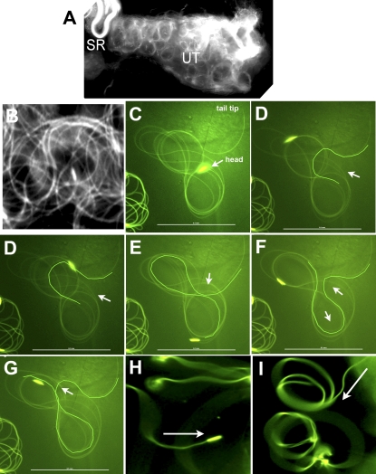
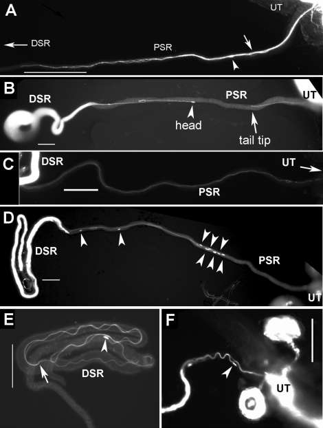
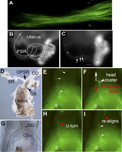
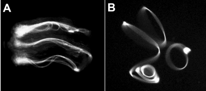
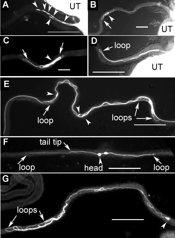
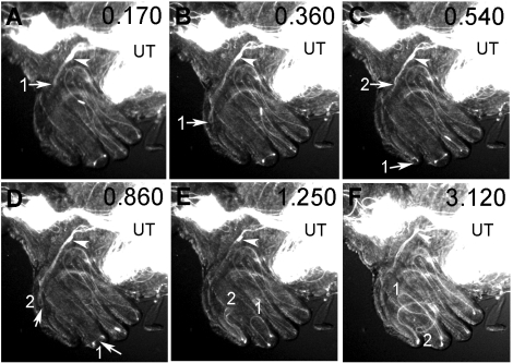
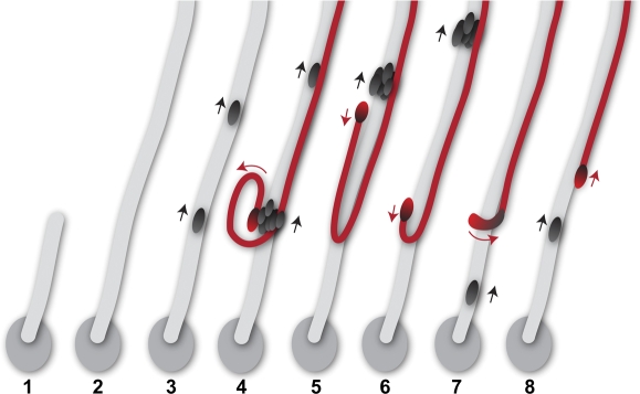
Motile cilia and flagella exhibit many waveforms as outputs of dynein activation sequences on the highly conserved axoneme. Motility change of sperm in the reproductive tract is difficult to study and remains an important area of investigation. Sperm typically execute a sinusoidal waveform. Increased viscosity in the medium induces somewhat unusual arc-line and helical waveforms in some sperm. However, whether the latter two waveforms occur in vivo is not known. Using green fluorescence protein imaging, we show that Drosophila sperm in the uterus move in circular foci via arc-line waves, predominantly in a tail-leading orientation. From the uterus, a small fraction of the sperm enters the seminal receptacle (SR) in parallel formations. After sperm storage and coincident with fertilization of the egg, the sperm exit the SR via head-leading helical waves. Consistent with the observed bidirectional movements, the sperm show the ability to propagate both base-to-tip and tip-to-base flagellar waves. Numerous studies have shown that sperm motility is regulated by intraflagellar calcium concentrations; in particular, the Pkd2 calcium channel has been shown to affect sperm storage. Our analyses here suggest that Pkd2 is required for the sperm to adopt the correct waveform and movement orientation during SR entry. A working model for the sperm's SR entry movement is proposed.


Figures

FIG. 1.
FIG. 1.
The arc-line, base-to-tip, and tip-to-base waves of sperm from the uterus. Snap views of live samples are shown. A) The moving sperm (∼5000 sperm) appear as circular foci in the uterus. B) A close-up view of the sperm in A shows circularly bent flagella (Supplemental Movies 1 and 2). CG) A sperm released from the uterus executes an arc-line waveform (Supplemental Movie 3). C) A single fly flagellum forms up to 10 arc-line bends that stack into a figure-8 configuration. DG) Sequential snap views showing formation of new arc-line bends (highlighted). Similar to sinusoidal waves, all new bends initiate at one of the flagellar ends (the tail end in this case). The arrow points to the thrust direction of a new bend that protrudes out as an open arc that then closes. The thrust direction alternates in opposite directions. The arc-line bends propagate through the entire folded flagellum, as evidenced by the moving head as it is continuously dragged behind its flagellum on the figure-8 path. H) Tip-to-base waves are observed when the distal flagellum is anchored (Supplemental Movie 4). I) Base-to-tip waves are observed when the proximal flagellum is anchored (Supplemental Movie 5). Arrows in H and I point to the direction of wave propagation. Bar = 0.1 mm.
FIG. 2.
FIG. 2.
Parallel formation of flagella during SR entry and exit. Still images from frozen samples are shown. AC) Images of sperm entry movement were obtained 15 min to 1 h after mating. A) Sperm entered the PSR in a parallel formation consisting of multiple sperm tails twisting around each other like a bundle. This tail bundle has entered halfway (0.5 mm) into the PSR. One head (arrowhead) and one flagellar loop (arrow), representing sperm with atypical flagellar configurations, were found in the mix of the tail bundle in A (Fig. 3F and Supplemental Movie 9). An explanation for these atypical sperm is given in Figure 8. B) Two sperm exhibit tail-leading PSR entry movement: One just entered the PSR, and the other nearly entered the DSR. The flagella of these sperm were in extended linear shape (a small fold was caused by unfolding the naturally folded PSR). C) Sperm moved across the PSR lumen without flagellar folds. D and F) Images of sperm exit movement were obtained 24 h after mating, and sperm exhibited head-leading movement (arrowheads indicate sperm heads). E) A sperm inside in the DSR is folded (arrow indicates, tail tip). Bar = 0.1 mm.
FIG. 3.
FIG. 3.
Leading tails and lagging heads. The dynamic process of PSR entry is shown here with video snap views of live samples. A) Multiple sperm tails enter the PSR in parallel formation like a bundle; the sperm are labeled with both head and tail GFP (Supplemental Movie 6). B and C) The sperm (labeled with head GFP only) pass through the convoluted PSR tubule (line trace in B) and enter the folded DSR on the right, which already contained a lot of sperm, as indicated by the bright fluorescence (Supplemental Movies 7 and 8). Arrows in C point to a cluster of three sperm heads that moved in one overlapping array consisting of eight sperm, which was then followed by a second array consisting of five sperm. The array is defined by having head-to-head spacing shorter than one sperm length of 2 mm (Supplemental Table S1). All of these sperm went through the PSR without to-and-fro motion and without turning or changing of the direction of movement. The time was 45 min after mating. D) Image of the uterus shows the relative locations of the oviduct (OD), ventral SR, and dorsal spermathecae (S). Arrows point to the openings of SR and spermathecae (OPSR and OS, respectively). G) Image at the uterus-oviduct junction provides spatial orientation for Supplemental Movie 9 (E, F, H, and I). Notice that the spermathecae are folded back onto the dorsal surface of the uterus. The dashed circle represents the location of the sperm focus, from which sperm initiate PSR entry movement in Supplemental Movie 9. The dashed arrow points to the direction of moving parallel sperm array. E, F, H, and I) Sequential snap views from Supplemental Movie 9 (time index, 1.375, 1.750, 3.375, and 4.375 sec). When multiple sperm flagella align into a parallel bundle, this appears as a GFP trail that leads upward toward the OPSR. E) Two sperm heads are on the GFP trail, and a head cluster is just about to be pulled out of the circular sperm focus in the uterus. F) The head cluster is pulled out and moving upward on the GFP trail. The head cluster contained one atypical sperm (red arrowhead), which is identifiable from its opposite moving direction and separation from the head cluster. The white arrow points to the upward-moving direction of most sperm, whereas the red arrow points to the downward-moving direction of the atypical sperm. H and I) The atypical sperm head undergoes a U-turn (H), and it rejoins the majority of sperm on the GFP trail (I). Original magnifications ×400 (A), ×20 (B, C), ×10 (D), ×40 (EI).
FIG. 4.
FIG. 4.
Arrangement of sperm bundle inside the PSR. AD) Still images of fast-frozen, wild-type sperm that entered the PSR. The uterus is positioned on the right beyond the view, and the DSR is on the left. A) This PSR is filled with sperm, with heads visible along the length (arrowheads). B) An enlarged image of A shows intertwining sperm flagellar bundles that are mostly parallel to each other. A small number of bent flagellar loops (arrows) are present in the mix. C) The sperm heads in the bundle are either well separated or clustered with a separation spacing of less than one head length (big arrowhead). D) Some clusters (arrowheads) contain as many as 22 sperm. E) The number of heads found in a cluster (x-axis) and observed frequency (y-axis) are plotted (n = 164 heads). Bar = 0.1 mm.
FIG. 5.
FIG. 5.
Sperm movement during and after storage in the DSR. Video snap views of live samples are shown. A) Several sperm move in folded forms in the DSR by protruding, hairpin-shaped flagellar bends (Supplemental Movie 10). The head is dragged backward by its flagellum as the flagellum slithers through its folded form. Occasionally, the head pauses and reverses to head-leading movement for a brief moment, resulting in a to-and-fro pattern of movement. B) This sperm released from the DSR is executing a helical waveform with head-leading clockwise rotations (Supplemental Movie 11). The sperm is free floating and not anchored at any points on the slide. Original magnification ×20.
FIG. 6.
FIG. 6.
Abnormal configurations of Pkd2 mutant sperm during PSR entry. Still images from frozen samples are shown. These are representative images of Pkd2-mutant sperm that moved into the PSR but appeared to be trapped. The uteri are positioned to the right. AD and EG show sperm from Pkd2KO67/Pkd2KO67 and Pkd21/Pkd2KO67 males, respectively. A) Six sperm moved into the PSR with head-leading (arrowheads) instead of tail-leading orientation observed for most wild-type sperm. B) The head of the sperm was leading, but flagellar loops (arrow) began to form. C) A sperm was folded excessively into an abnormal yarn ball appearance. D) A hairpin-shaped flagellar loop entered the PSR (Supplemental Movie 12). E) Several sperm were tangled together and contained multiple figure-8-shaped loops (arrows). F) A figure-8-shaped flagellum was located within PSR lumen. G) A tail-leading sperm was found to form multiple flagellar buckles. Bar = 0.1 mm.
FIG. 7.
FIG. 7.
Abnormal propulsion of Pkd2-mutant flagellum during PSR entry. Sequential video snap views from Supplemental Movie 12 are used to show the abnormal propulsion of the mutant sperm at the uterus-PSR junction (AF). To orient viewers, the PSR tubule forms the left outer rim of the coiled SR complex, going underneath the DSR tubules that face the reader. The sperm head (arrowhead) stayed in the general vicinity while its flagellum was protruding two hairpin-shaped flagellar loops (labeled 1 and 2) into the PSR lumen. The “hairpins” soon became stuck even though the flagellum continued to beat. The head was pulled along but stayed in the general area. The time index (sec) is shown at top right corner of each frame. Original magnification ×20.
FIG. 8.
FIG. 8.
A working model for PSR entry. Key events are shown in sequence. 1) Sperm move in circular foci in the uterus. One of the foci is depicted as an oval, representing the focus that has responded to the PSR entry signal. From this, sperm begin to form a parallel array with tails leading (gray stripe). 2) The sperm in the parallel array move into the PSR lumen on the top beyond our view, leading to the appearance of a GFP trail in Supplemental Movie 9. 3) As the sperm bundle moves upward, additional sperm are added from the circular focus, and the heads of these sperm appear on the trail at different times and move upward. This represents the behavior of most sperm entering the PSR (black heads). 4) A cluster of sperm heads, including one misaligned atypical head (red), appears on the trail and moves upward. 5 and 6) The atypical head is recognized by its subsequent separation from the head cluster and movement in an opposite direction (red arrow) with respect to the majority of sperm on the trail. If the situation depicted in 5 occurs within the PSR lumen, it would give rise to one head plus one flagellar loop in the mix of a tail bundle, as shown in Figure 2A. 6) As the tail of the atypical sperm continues to move upward, the head makes a U-turn and subsequently moves upward, just like the majority of sperm on the trail.

Similar articles

Cited by

References

    1. Eisenbach M, Giojalas LC. Sperm guidance in mammals—an unpaved road to the egg. Nat Rev Mol Cell Biol 2006; 7: 276 285 - PubMed
    1. Suarez SS. Regulation of sperm storage and movement in the mammalian oviduct. Int J Dev Biol 2008; 52: 455 462 - PubMed
    1. Cho C, Bunch DO, Faure JE, Goulding EH, Eddy EM, Primakoff P, Myles DG. Fertilization defects in sperm from mice lacking fertilin beta. Science 1998; 281: 1857 1859 - PubMed
    1. Ikawa M, Nakanishi T, Yamada S, Wada I, Kominami K, Tanaka H, Nozaki M, Nishimune Y, Okabe M. Calmegin is required for fertilin alpha/beta heterodimerization and sperm fertility. Dev Biol 2001; 240: 254 261 - PubMed
    1. Hagaman JR, Moyer JS, Bachman ES, Sibony M, Magyar PL, Welch JE, Smithies O, Krege JH, O'Brien DA. Angiotensin-converting enzyme and male fertility. Proc Natl Acad Sci U S A 1998; 95: 2552 2557 - PMC - PubMed

Publication types

MeSH terms