. 2011 Feb;121(2):703-14.
doi: 10.1172/JCI44740.

Deficiency of liver sinusoidal scavenger receptors stabilin-1 and -2 in mice causes glomerulofibrotic nephropathy via impaired hepatic clearance of noxious blood factors

Affiliations

Deficiency of liver sinusoidal scavenger receptors stabilin-1 and -2 in mice causes glomerulofibrotic nephropathy via impaired hepatic clearance of noxious blood factors

Kai Schledzewski et al. J Clin Invest. 2011 Feb.

Abstract

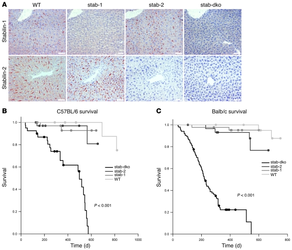
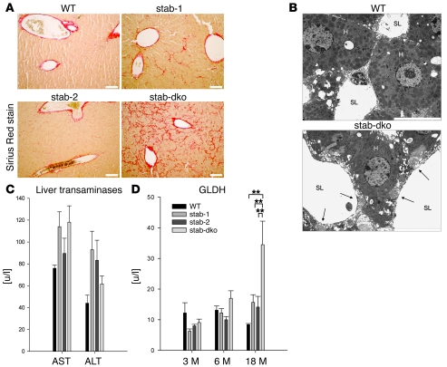
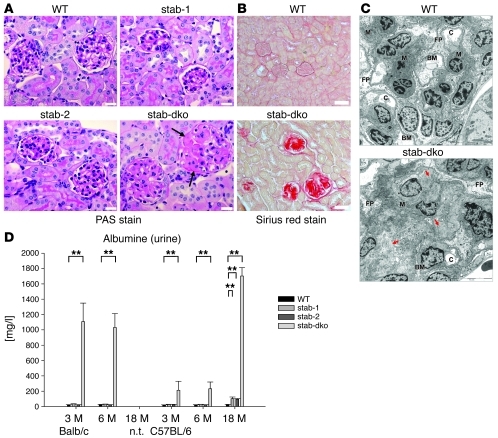
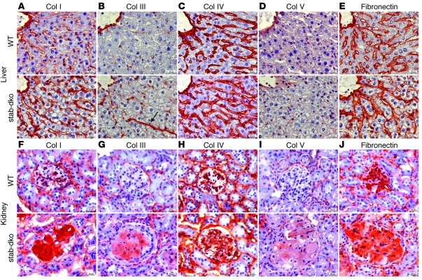
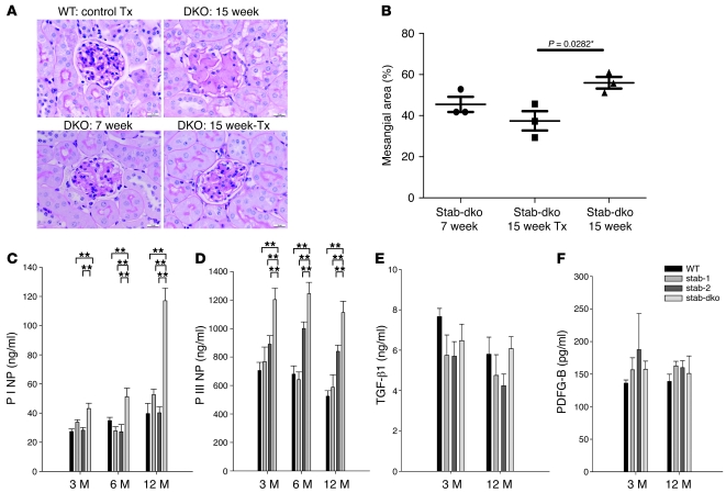
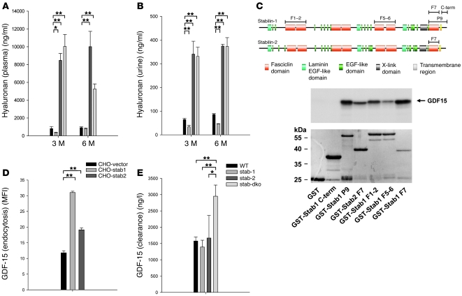
Tissue homeostasis and remodeling are processes that involve high turnover of biological macromolecules. Many of the waste molecules that are by-products or degradation intermediates of biological macromolecule turnover enter the circulation and are subsequently cleared by liver sinusoidal endothelial cells (LSEC). Besides the mannose receptor, stabilin-1 and stabilin-2 are the major scavenger receptors expressed by LSEC. To more clearly elucidate the functions of stabilin-1 and -2, we have generated mice lacking stabilin-1, stabilin-2, or both stabilin-1 and -2 (Stab1–/– Stab2–/– mice). Mice lacking either stabilin-1 or stabilin-2 were phenotypically normal; however, Stab1–/– Stab2–/– mice exhibited premature mortality and developed severe glomerular fibrosis, while the liver showed only mild perisinusoidal fibrosis without dysfunction. Upon kidney transplantation into WT mice, progression of glomerular fibrosis was halted, indicating the presence of profibrotic factors in the circulation of Stab1–/– Stab2–/– mice. While plasma levels of known profibrotic cytokines were unaltered, clearance of the TGF-β family member growth differentiation factor 15 (GDF-15) was markedly impaired in Stab1–/– Stab2–/– mice but not in either Stab1–/– or Stab2–/– mice, indicating that it is a common ligand of both stabilin-1 and stabilin-2. These data lead us to conclude that stabilin-1 and -2 together guarantee proper hepatic clearance of potentially noxious agents in the blood and maintain tissue homeostasis not only in the liver but also distant organs.


Figures

Figure 1
Figure 1. Generation of Stab1–/–, Stab2–/–, and Stab1–/–Stab2–/– mice.
(A) Representative liver sections of 3-month-old WT, Stab1–/– (stab-1), Stab2–/– (stab-2), and Stab1–/–Stab2–/– (stab-dko) mice of C57BL/6 genetic background were stained by immunohistochemistry with antibodies against stabilin-1 (upper row) and stabilin-2 (lower row). WT sections show sinusoidal distribution of both stabilins. No stabilin-1 was detected in Stab1–/–, no stabilin-2 was detected in Stab2–/–, and neither of the stabilins were expressed in Stab1–/–Stab2–/– animals. Original magnification, ×200. Scale bars: 50 μm. (B) Survival of WT, Stab1–/–, Stab2–/–, and Stab1–/–Stab2–/– mice (C57BL/6 background) over a period of 820 days (n = 38, n = 45, n = 43, and n = 40, respectively). Mean survival of Stab1–/–Stab2–/– mice was significantly (P < 0.001) reduced (415 d), if compared with WT (803 d), Stab1–/– (615 d), and Stab2–/– (625 d) strains. Single-deficient mice displayed a tendency toward reduced lifespan (Stab1–/– [P = 0.13] and Stab2–/– [P = 0.12]). (C) Survival of WT, Stab1–/–, Stab2–/–, and Stab1–/–Stab2–/– mice of Balb/c genetic background over a period of 751 days (n = 31, n = 44, n = 30, and n = 102, respectively). Mean survival of Stab1–/–Stab2–/– mice was dramatically reduced (269 d), if compared with WT (739 d), Stab1–/– (584 d), and Stab2–/– (618 d). Single-deficient mice displayed a tendency toward a reduced lifespan (Stab1–/– [P = 0.28]) and Stab2–/– [P = 0.06]) that did not reach the level of confidence (P < 0.05). (B and C) The survival data were analyzed by log-rank statistics, and pair-wise multiple comparison procedures were done by the Holm-Sidak method.
Figure 2
Figure 2. Liver phenotype of stabilin-deficient mice.
(A) Representative liver sections of 6-month-old WT, Stab1–/–, Stab2–/–, and Stab1–/–Stab2–/– mice of C57BL/6 genetic background were stained with Sirius red to identify the degree of fibrosis (n = 5). No Sirius red–positive fibers except around large blood vessels were detected in WT specimens, whereas few positive fibers were seen in Stab1–/– and Stab2–/– specimens. Sirius red staining of Stab1–/–Stab2–/– livers identified fibrotic fibers along the sinusoidal network. Original magnification, ×100. Scale bars: 100 μm. (B) Representative transmission EM images of liver sinusoids captured from 6-month-old WT as well as Stab1–/–Stab2–/– mice. Collagen fibers in the space of Disse close to the sinusoidal lumen are indicated by arrows. H, hepatocyte; SL, sinusoidal lumen. n = 3. Original magnification, ×10,000. Scale bar: 10 μm. (C) Activity levels of liver AST and ALT were measured in the plasma of 18-month-old C57BL/6 WT, C57BL/6 Stab1–/–, C57BL/6 Stab2–/–, and C57BL/6 Stab1–/–Stab2–/– mice. No statistically significant differences were found, indicating normal liver function even in very old Stab1–/–Stab2–/– animals. Data are mean ± SEM from 5 mice. (D) Activity levels of GLDH were measured in the plasma of C57BL/6 WT, C57BL/6 Stab1–/–, C57BL/6 Stab2–/–, and C57BL/6 Stab1–/–Stab2–/– mice of the indicated age as a marker of necrotic processes. A significant increase of GLDH was observed only in 18-month-old C57BL/6 animals. Data represent mean ± SEM from 5 mice. **P < 0.01 relative to Stab1–/–Stab2–/– values.
Figure 3
Figure 3. Kidney phenotype of stabilin-deficient mice.
(A) Representative PAS-stained kidney sections from 3-month-old WT, Stab1–/–, Stab2–/–, and Stab1–/–Stab2–/– mice of Balb/c genetic background. The glomeruli from WT as well as Stab1–/– and Stab2–/– mice had a normal appearance. In contrast, mesangial expansion was evident in Stab1–/–Stab2–/– mice (arrows) (n = 3). Original magnification, ×400. Scale bars: 20 μm. (B) Representative kidney sections of 3-month-old WT and Stab1–/–Stab2–/– mice of C57BL/6 background were stained with Sirius red to identify the degree of fibrosis (n = 3). Strong Sirius red–positive staining indicating collagen deposition was detected only in Stab1–/–Stab2–/– glomeruli. The extent of the glomerulosclerosis was similar in Stab1–/–Stab2–/– mice of both Balb/c and C57BL/6 genetic background (not shown). Original magnification, ×200. Scale bars: 50 μm. (C) Representative transmission EM images of 4-week-old Stab1–/–Stab2–/– glomeruli of Balb/c genetic background as compared with WT showed large deposits of fibrillar material (red arrows) and a severely enlarged mesangium (M) with narrowing of capillary lumens (C), partial loss of foot processes (FP) and regular basement membrane (BM). n = 3. Original magnification, ×10,000. Scale bars: 10 μm. (D) Urine of 3- and 6-month-old Balb/c and 3-, 6-, and 18-month-old C57BL/6 mice of all genotypes was collected and analyzed for albumin content by competitive ELISA. Albumine levels were significantly increased in all Stab1–/–Stab2–/– samples in comparison with WT and single-deficient animals. Data represent mean ± SEM from 5 mice. **P < 0.01 relative to WT values.
Figure 4
Figure 4. Analysis of extracellular matrix content in liver and kidney of stabilin-deficient mice.
(AE) Representative liver sections of 3-month-old C57BL/6 WT (first row) and C57BL/6 Stab1–/–Stab2–/– mice (second row) were stained by immunohistochemistry with antibodies against (A) collagen I, (B) collagen III, (C) collagen IV, (D) collagen V, and (E) fibronectin. Similar to the findings in Sirius red–stained samples, collagen III fibers (B, arrow) occurred along the sinusoidal network. (FJ) Representative kidney sections of 3-month-old C57BL/6 WT (third row) and C57BL/6 Stab1–/–Stab2–/– mice (fourth row) were stained by immunohistochemistry with antibodies against (F) collagen I (G), collagen III (H), collagen IV (I), collagen V, and (J) fibronectin. (AJ) The results of the immunohistochemical experiments are described in detail in Results. Original magnification, ×400. Scale bars: 20 μm.
Figure 5
Figure 5. Effect of circulating factors on kidney phenotype.
(A) Representative PAS-stained sections of kidneys from 7-week-old (lower left) and 15-week-old (upper right) Stab1–/–Stab2–/– mice and of Stab1–/–Stab2–/––to–WT (lower right) as well as WT-to-WT (upper left) kidney transplants at 56 days after transplantation are shown (n = 3). While WT control transplants (Tx) were unaffected and Stab1–/–Stab2–/– glomeruli of 15-week-old animals displayed progression of glomerular fibrosis, glomeruli of Stab1–/–Stab2–/––to–WT renal transplants showed regression of fibrosis after transplantation. Original magnification, ×400. (B) Quantitative evaluation of glomerular fibrosis in Stab1–/–Stab2–/––to–WT renal transplants after transplantation (n = 3) showed a statistically significant decrease in mesangial area (P = 0.0282) as compared with 15-week-old Stab1–/–Stab2–/– kidneys, while comparison with 7-week-old Stab1–/–Stab2–/– kidneys showed a tendency toward reduction of mesangial area. (C and D) Plasma samples of 3-, 6-, and 12-month-old WT, Stab1–/–, Stab2–/–, and Stab1–/–Stab2–/– mice of C57BL/6 genetic background were analyzed for P I NP and P III N) by ELISA. As only stabilin-2, but not stabilin-1, is a scavenger receptor for these propeptides, elevated levels of the propeptides found only in Stab1–/–Stab2–/– animals indicate increased de novo synthesis of collagens in Stab1–/–Stab2–/– mice. (E and F) Plasma samples of 3- and 12-month-old WT, Stab1–/–, Stab2–/–, and Stab1–/–Stab2–/– mice (C57BL/6 background) were measured for TGF-β1 (E) and PDFG-B (F) by ELISA. No elevated levels of known profibrotic cytokines were detected in the tested stabilin animals Data represent mean ± SEM for at least 5 mice per genotype. **P < 0.01.
Figure 6
Figure 6. Differential impairment of hepatic blood clearance for stabilin ligands.
(A and B) Plasma (A) and urine (B) HA levels of 3- and 6-month-old WT, Stab1–/–, Stab2–/–, and Stab1–/–Stab2–/– mice of C57BL/6 genetic background were measured by ELISA. Elevated levels of HA were detected exclusively in Stab2–/– and Stab1–/–Stab2–/– animals, but not in Stab1–/– mice. Data represent mean ± SEM for at least 5 mice per genotype. (C) Schematic presentation of the protein domain organization of stabilin-1 and stabilin-2 shows the fragments used in the yeast 2-hybrid screening (P9) and GST-pull down assays. Stabilin-1 F1-2, stabilin-1 F5-6, stabilin-1 F7, stabilin-1 P9, stabilin-1 cytoplasmic tail (C-term), and stabilin-2 F7 are indicated by bars. GST pull-down assay demonstrating that fasciclin domains of both stabilin-1 and stabilin-2 interact with in vitro–translated GDF-15. GDF-15 neither binds to GST alone (negative control) nor to the GST-fused cytoplasmic tail of stabilin-1. n = 3. (D) Quantification of stabilin-1– and stabilin-2–mediated uptake of Alexa Fluor 488–labeled GDF-15 using flow cytometry. n = 3. Data represent mean ± SEM for 3 independent experiments. (E) 5 μg of recombinant human GDF-15 was injected in the tail vein of WT, Stab1–/–, Stab2, and Stab1–/–Stab2–/– mice of Balb/c genetic background, and plasma levels of hGDF-15 were measured after 5 hours (n = 16, n = 3, n = 3, n = 12, respectively). Increased levels of hGDF-15 were exclusively observed in Stab1–/–Stab2–/– samples indicating impaired clearance of GDF-15 only upon combined absence of both stabilin receptors. Data represent mean ± SEM. *P < 0.05; **P < 0.01.

Similar articles

Cited by

References

    1. Smedsrod B. Clearance function of scavenger endothelial cells. Comp Hepatol. 2004;3 suppl 1:S22. - PMC - PubMed
    1. Seternes T, Sorensen K, Smedsrod B. Scavenger endothelial cells of vertebrates: a nonperipheral leukocyte system for high-capacity elimination of waste macromolecules. Proc Natl Acad Sci U S A. 2002;99(11):7594–7597. doi: 10.1073/pnas.102173299. - DOI - PMC - PubMed
    1. Malovic I, et al. The mannose receptor on murine liver sinusoidal endothelial cells is the main denatured collagen clearance receptor. Hepatology. 2007;45(6):1454–1461. doi: 10.1002/hep.21639. - DOI - PubMed
    1. Elvevold K, Simon-Santamaria J, Hasvold H, McCourt P, Smedsrod B, Sorensen KK. Liver sinusoidal endothelial cells depend on mannose receptor-mediated recruitment of lysosomal enzymes for normal degradation capacity. Hepatology. 2008;48(6):2007–2015. doi: 10.1002/hep.22527. - DOI - PubMed
    1. Muro H, Shirasawa H, Kosugi I, Nakamura S. Defect of Fc receptors and phenotypical changes in sinusoidal endothelial cells in human liver cirrhosis. . Am J Pathol. 1993;143(1):105–120. - PMC - PubMed

Publication types