

Adrenal hemangioma in a 19-year-old female
- PMID: 21293064
- PMCID: PMC3156522
- DOI: 10.4103/0256-4947.76411
Adrenal hemangioma in a 19-year-old female
Abstract
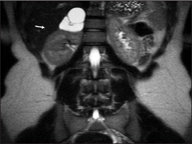
Adrenal masses are being detected with increasing frequency due to the widespread use of computed tomography, magnetic resonance imaging and even ultrasonography for the evaluation of diseases with abdominal involvement. It is estimated that adrenal masses are an accidental finding in 1% to 5% of all abdominal CT scans performed. Adrenal hemangiomas are rare and nonfunctioning benign tumors and their differential diagnosis preoperatively is rather challenging. Adrenal hemangiomas are most usually cavernous, unilateral lesions of the adrenal glands; bilateral involvement has been reported twice, which appears between the ages 50 and 70 years, with a 2:1 female-to-male ratio. Approximately 60 surgical cases have been reported in the literature. In general, this tumor is large, and all cases reported were treated with open surgery or retroperitoneoscopic procedure. We report a case of a 19-year-old female patient with adrenal hemangioma that was removed by laparoscopic adrenalectomy.
Figures


References
-
- Del Gaudio A, Solidoro G, Martinelli G. Adrenal hemangiomas: Two case reports with a review of the literature. Surgery. 1989;105:674–81. - PubMed
-
- Honig SC, Klavans MS, Hyde C, Siroky MB. Adrenal hemangioma: An unusual adrenal mass delineated with magnetic resonance imaging. J Urol. 1991;146:400–2. - PubMed
-
- Salup R, Finegold R, Borochovitz D, Boehnke M, Posner M. Cavernous hemangioma of the adrenal gland. J Urol. 1992;147:110–2. - PubMed
-
- Carbonell LC, Toro AO, Llanes VJ, Gali BO, Mas GA. Adrenal hemangioma: Review of the literature. Prog Urol. 1996;6:292–6. - PubMed
-
- Oh BR, Jeong YY, Ryu SB, Park YI, Kang HK. A case of adrenal cavernous hemangioma. Int J Urol. 1997;4:608–10. - PubMed
Publication types
MeSH terms
LinkOut - more resources
Full Text Sources
Medical