

Infrared scanning laser ophthalmoscope imaging of the macula and its correlation with functional loss and structural changes in patients with stargardt disease
- PMID: 21293320
- PMCID: PMC3116073
- DOI: 10.1097/IAE.0b013e3181f441f6
Infrared scanning laser ophthalmoscope imaging of the macula and its correlation with functional loss and structural changes in patients with stargardt disease
Abstract
Purpose: To correlate the degree of functional loss with structural changes in patients with Stargardt disease.
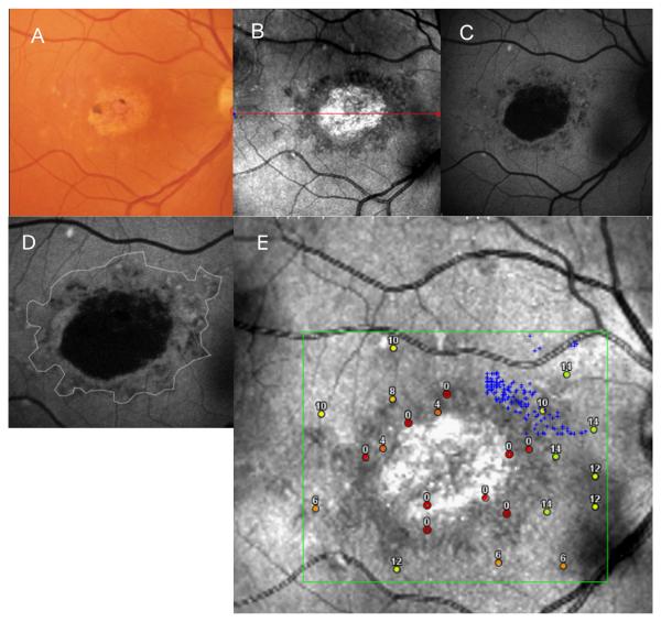
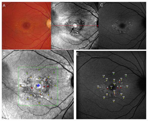
Methods: Eighteen eyes of 10 patients with Stargardt disease were studied. Scanning laser ophthalmoscope infrared images were compared with corresponding spectral-domain optical coherence tomography scans. Additionally, scanning laser ophthalmoscope microperimetry was performed, and results were superimposed on scanning laser ophthalmoscope infrared images and in selected cases on fundus autofluorescence images.
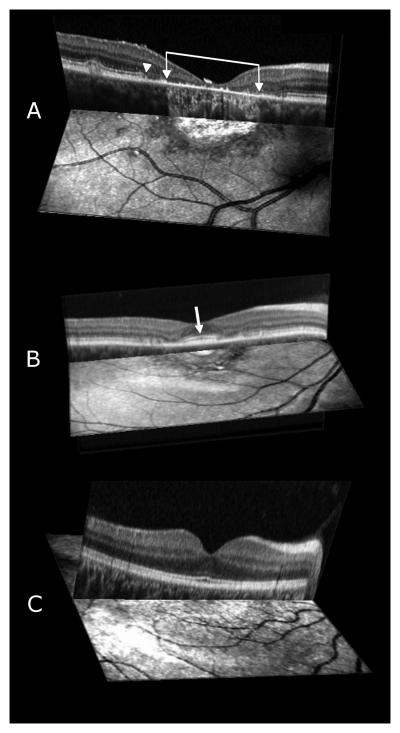
Results: Seventeen of 18 eyes showed a distinct hyporeflective foveal and/or perifoveal area with distinct borders on scanning laser ophthalmoscope infrared images, which was less evident on funduscopy and incompletely depicted in fundus autofluorescence images. This hyporeflective zone corresponded to areas of significantly elevated psychophysical thresholds on microperimetry testing, in addition to thinning of the retinal pigment epithelium and disorganization or loss of the photoreceptor cell inner segment-outer segment junction and external-limiting membrane on spectral-domain optical coherence tomography.
Conclusion: Scanning laser ophthalmoscope infrared fundus images are useful for depicting retinal structural changes in patients with Stargardt disease. A spectral-domain optical coherence tomography/scanning laser ophthalmoscope microperimetry device allows for a direct correlation of structural abnormalities with functional defects that will likely be applicable for the determination of retinal areas for potential improvement of retinal function in these patients during future clinical trials and for the monitoring of the diseases' natural history.
Figures
Similar articles
-
Association of dark-adapted visual function with retinal structural changes in patients with Stargardt disease.Retina. 2014 May;34(5):989-95. doi: 10.1097/IAE.0000000000000022. Retina. 2014. PMID: 24280667 Free PMC article.
-
Correlation between photoreceptor layer integrity and visual function in patients with Stargardt disease: implications for gene therapy.Invest Ophthalmol Vis Sci. 2012 Jul 3;53(8):4409-15. doi: 10.1167/iovs.11-8201. Invest Ophthalmol Vis Sci. 2012. PMID: 22661472 Free PMC article.
-
The value of retinal imaging with infrared scanning laser ophthalmoscopy in patients with stargardt disease.Retina. 2014 Jul;34(7):1391-9. doi: 10.1097/IAE.0000000000000070. Retina. 2014. PMID: 24317291 Free PMC article.
-
[Pathophysiology of macular diseases--morphology and function].Nippon Ganka Gakkai Zasshi. 2011 Mar;115(3):238-74; discussion 275. Nippon Ganka Gakkai Zasshi. 2011. PMID: 21476310 Review. Japanese.
-
Macular telangiectasia type 2.Prog Retin Eye Res. 2013 May;34:49-77. doi: 10.1016/j.preteyeres.2012.11.002. Epub 2012 Dec 3. Prog Retin Eye Res. 2013. PMID: 23219692 Free PMC article. Review.
Cited by
-
Inner and outer retinal changes in retinal degenerations associated with ABCA4 mutations.Invest Ophthalmol Vis Sci. 2014 Mar 20;55(3):1810-22. doi: 10.1167/iovs.13-13768. Invest Ophthalmol Vis Sci. 2014. PMID: 24550365 Free PMC article.
-
Multimodal fundus imaging in fundus albipunctatus with RDH5 mutation: a newly identified compound heterozygous mutation and review of the literature.Doc Ophthalmol. 2012 Aug;125(1):51-62. doi: 10.1007/s10633-012-9336-z. Epub 2012 Jun 6. Doc Ophthalmol. 2012. PMID: 22669287 Review.
-
Perifoveal Cone- and Rod-Mediated Temporal Contrast Sensitivities in Stargardt Disease/Fundus Flavimaculatus.Invest Ophthalmol Vis Sci. 2021 Nov 1;62(14):24. doi: 10.1167/iovs.62.14.24. Invest Ophthalmol Vis Sci. 2021. PMID: 34807235 Free PMC article.
-
Transition zones between healthy and diseased retina in choroideremia (CHM) and Stargardt disease (STGD) as compared to retinitis pigmentosa (RP).Invest Ophthalmol Vis Sci. 2011 Dec 20;52(13):9581-90. doi: 10.1167/iovs.11-8554. Invest Ophthalmol Vis Sci. 2011. PMID: 22076985 Free PMC article.
-
The role of multimodal imaging and vision function testing in ABCA4-related retinopathies and their relevance to future therapeutic interventions.Ther Adv Ophthalmol. 2021 Dec 19;13:25158414211056384. doi: 10.1177/25158414211056384. eCollection 2021 Jan-Dec. Ther Adv Ophthalmol. 2021. PMID: 34988368 Free PMC article. Review.
References
-
- Stargardt K. Uber familiare progressive degeneration in der makulagegend des auges. Albrecht von Graefes Arch Klin Ophthalmol. 1909;71:534–550.
-
- Blacharski PA. Fundus flavimaculatus. In: Newsome DA, editor. Retinal dystrophies and Degenerations. Raven Press; New York: 1988. pp. 135–159.
-
- Westerfeld C, Mukai S. Stargardt's disease and the ABCA4 gene. Seminars Ophthalmol. 2008;23:59–65. - PubMed
-
- Fishman GA. Fundus flavimaculatus. A clinical classification. Arch Ophthalmol. 1976;94:2061–2067. - PubMed
-
- Ergun E, Hermann B, Wirtitsch M, et al. Assessment of central visual function in Stargardt Disease/Fundus Flavimaculatus with Ultrahigh-Resolution Optical Coherence Tomography. Invest Ophthalmol & Vis Sci. 2005;46:310–316. - PubMed
Publication types
MeSH terms
Substances
Grants and funding
LinkOut - more resources
Full Text Sources
Other Literature Sources
Medical
