. 2011 Feb 3;470(7332):73-7.
doi: 10.1038/nature09750.

Femtosecond X-ray protein nanocrystallography

Henry N Chapman  1 Petra FrommeAnton BartyThomas A WhiteRichard A KirianAndrew AquilaMark S HunterJoachim SchulzDaniel P DePonteUwe WeierstallR Bruce DoakFilipe R N C MaiaAndrew V MartinIlme SchlichtingLukas LombNicola CoppolaRobert L ShoemanSascha W EppRobert HartmannDaniel RollesArtem RudenkoLutz FoucarNils KimmelGeorg WeidenspointnerPeter HollMengning LiangMiriam BarthelmessCarl CalemanSébastien BoutetMichael J BoganJacek KrzywinskiChristoph BostedtSaša BajtLars GumprechtBenedikt RudekBenjamin ErkCarlo SchmidtAndré HömkeChristian ReichDaniel PietschnerLothar StrüderGünter HauserHubert GorkeJoachim UllrichSven HerrmannGerhard SchallerFlorian SchopperHeike SoltauKai-Uwe KühnelMarc MesserschmidtJohn D BozekStefan P Hau-RiegeMatthias FrankChristina Y HamptonRaymond G SierraDmitri StarodubGarth J WilliamsJanos HajduNicusor TimneanuM Marvin SeibertJakob AndreassonAndrea RockerOlof JönssonMartin SvendaStephan SternKarol NassRobert AndritschkeClaus-Dieter SchröterFaton KrasniqiMario BottKevin E SchmidtXiaoyu WangIngo GrotjohannJames M HoltonThomas R M BarendsRichard NeutzeStefano MarchesiniRaimund FrommeSebastian SchorbDaniela RuppMarcus AdolphTais GorkhoverInger AnderssonHelmut HirsemannGuillaume PotdevinHeinz GraafsmaBjörn NilssonJohn C H Spence
Affiliations

Femtosecond X-ray protein nanocrystallography

Henry N Chapman et al. Nature. .

Abstract

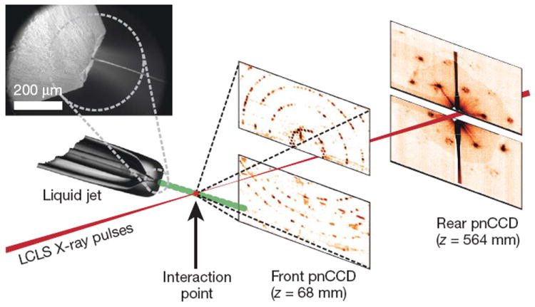
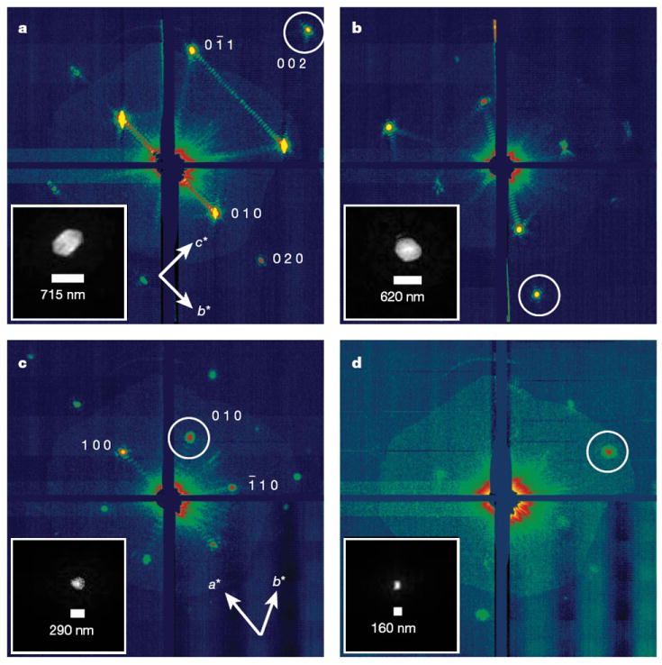
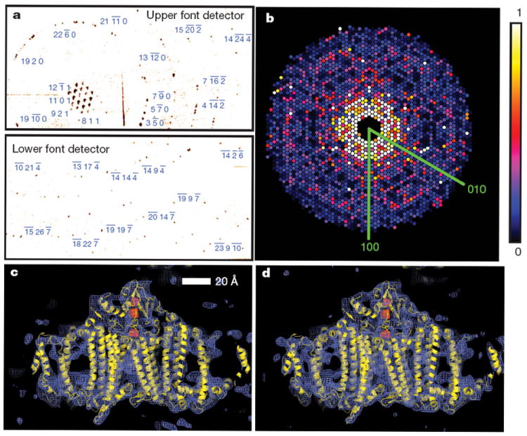
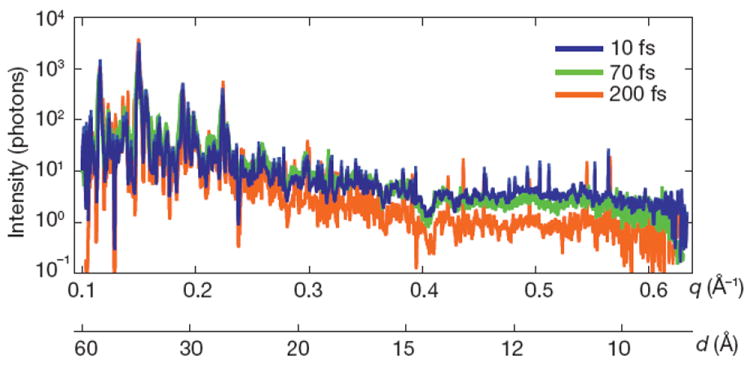
X-ray crystallography provides the vast majority of macromolecular structures, but the success of the method relies on growing crystals of sufficient size. In conventional measurements, the necessary increase in X-ray dose to record data from crystals that are too small leads to extensive damage before a diffraction signal can be recorded. It is particularly challenging to obtain large, well-diffracting crystals of membrane proteins, for which fewer than 300 unique structures have been determined despite their importance in all living cells. Here we present a method for structure determination where single-crystal X-ray diffraction 'snapshots' are collected from a fully hydrated stream of nanocrystals using femtosecond pulses from a hard-X-ray free-electron laser, the Linac Coherent Light Source. We prove this concept with nanocrystals of photosystem I, one of the largest membrane protein complexes. More than 3,000,000 diffraction patterns were collected in this study, and a three-dimensional data set was assembled from individual photosystem I nanocrystals (∼200 nm to 2 μm in size). We mitigate the problem of radiation damage in crystallography by using pulses briefer than the timescale of most damage processes. This offers a new approach to structure determination of macromolecules that do not yield crystals of sufficient size for studies using conventional radiation sources or are particularly sensitive to radiation damage.


Conflict of interest statement

The authors declare no competing financial interests.

Figures

Figure 1
Figure 1. Femtosecond nanocrystallography
Nanocrystals flow in their buffer solution in a gas-focused, 4-μm-diameter jet at a velocity of 10 m s−1 perpendicular to the pulsed X-ray FEL beam that is focused on the jet. Inset, environmental scanning electron micrograph of the nozzle, flowing jet and focusing gas. Two pairs of high-frame-rate pnCCD detectors record low-and high-angle diffraction from single X-ray FEL pulses, at the FEL repetition rate of 30 Hz. Crystals arrive at random times and orientations in the beam, and the probability of hitting one is proportional to the crystal concentration.
Figure 2
Figure 2. Coherent crystal diffraction
Low-angle diffraction patterns recorded on the rear pnCCDs, revealing coherent diffraction from the structure of the photosystem I nanocrystals, shown using a logarithmic, false-colour scale. The Miller indices of the peaks in a were identified from the corresponding high-angle pattern. In c we count seven fringes in the b* direction, corresponding to nine unit cells, or 250 nm. Insets, real-space images of the nanocrystal, determined by phase retrieval (using the Shrinkwrap algorithm) of the circled coherent Bragg shape transform.
Figure 3
Figure 3. Diffraction intensities and electron density of photosystem I
a, Diffraction pattern recorded on the front pnCCDs with a single 70-fs pulse after background subtraction and correction of saturated pixels. Some peaks are labelled with their Miller indices. The resolution in the lower detector corner is 8.5 Å. b, Precession-style pattern of the [001] zone for photosystem I, obtained from merging femtosecond nanocrystal data from over 15,000 nanocrystal patterns, displayed on the linear colour scale shown on the right. c, d, Region of the 2mFoDFc electron density map at 1.0σ (purple mesh), calculated from the 70-fs data (c) and from conventional synchrotron data truncated at a resolution of 8.5 Å and collected at a temperature of 100 K (d) (Methods). The refined model is depicted in yellow.
Figure 4
Figure 4. Pulse-duration dependence of diffraction intensities
Plot of the integrated Bragg intensities of photosystem I nanocrystal diffraction as a function of photon momentum transfer, q = (4π/λ)sin(θ) = 2π/d (wavelength, λ; scattering angle 2θ; resolution, d) for pulse durations of 10, 70 and 200 fs. Averages were obtained by isolating Bragg spots from 97,883, 805,311 and 66,063 patterns, respectively, normalized to pulse fluence. The error in each plot is indicated by the thickness of the line. The decrease in irradiance for 200-fs pulses and d < 25 Å indicates radiation damage for these long pulses, which is not apparent for 70-fs pulses and shorter.

Comment in

  • Diffraction before destruction.
    Doerr A. Doerr A. Nat Methods. 2011 Apr;8(4):283. doi: 10.1038/nmeth0411-283. Nat Methods. 2011. PMID: 21574275 No abstract available.

Similar articles

  • Toward structure determination using membrane-protein nanocrystals and microcrystals.
    Hunter MS, Fromme P. Hunter MS, et al. Methods. 2011 Dec;55(4):387-404. doi: 10.1016/j.ymeth.2011.12.006. Epub 2011 Dec 22. Methods. 2011. PMID: 22197730 Free PMC article. Review.
  • Femtosecond nanocrystallography using X-ray lasers for membrane protein structure determination.
    Fromme P, Spence JC. Fromme P, et al. Curr Opin Struct Biol. 2011 Aug;21(4):509-16. doi: 10.1016/j.sbi.2011.06.001. Curr Opin Struct Biol. 2011. PMID: 21752635 Free PMC article. Review.
  • Continuous X-ray diffractive field in protein nanocrystallography.
    Dilanian RA, Streltsov VA, Quiney HM, Nugent KA. Dilanian RA, et al. Acta Crystallogr A. 2013 Jan;69(Pt 1):108-18. doi: 10.1107/S0108767312042535. Epub 2012 Nov 16. Acta Crystallogr A. 2013. PMID: 23250067
  • Time-resolved protein nanocrystallography using an X-ray free-electron laser.
    Aquila A, Hunter MS, Doak RB, Kirian RA, Fromme P, White TA, Andreasson J, Arnlund D, Bajt S, Barends TR, Barthelmess M, Bogan MJ, Bostedt C, Bottin H, Bozek JD, Caleman C, Coppola N, Davidsson J, DePonte DP, Elser V, Epp SW, Erk B, Fleckenstein H, Foucar L, Frank M, Fromme R, Graafsma H, Grotjohann I, Gumprecht L, Hajdu J, Hampton CY, Hartmann A, Hartmann R, Hau-Riege S, Hauser G, Hirsemann H, Holl P, Holton JM, Hömke A, Johansson L, Kimmel N, Kassemeyer S, Krasniqi F, Kühnel KU, Liang M, Lomb L, Malmerberg E, Marchesini S, Martin AV, Maia FR, Messerschmidt M, Nass K, Reich C, Neutze R, Rolles D, Rudek B, Rudenko A, Schlichting I, Schmidt C, Schmidt KE, Schulz J, Seibert MM, Shoeman RL, Sierra R, Soltau H, Starodub D, Stellato F, Stern S, Strüder L, Timneanu N, Ullrich J, Wang X, Williams GJ, Weidenspointner G, Weierstall U, Wunderer C, Barty A, Spence JC, Chapman HN. Aquila A, et al. Opt Express. 2012 Jan 30;20(3):2706-16. doi: 10.1364/OE.20.002706. Opt Express. 2012. PMID: 22330507 Free PMC article.
  • X-ray diffraction from membrane protein nanocrystals.
    Hunter MS, DePonte DP, Shapiro DA, Kirian RA, Wang X, Starodub D, Marchesini S, Weierstall U, Doak RB, Spence JC, Fromme P. Hunter MS, et al. Biophys J. 2011 Jan 5;100(1):198-206. doi: 10.1016/j.bpj.2010.10.049. Biophys J. 2011. PMID: 21190672 Free PMC article.

Cited by

References

    1. Owen RL, Rudino-Pinera E, Garman EF. Experimental determination of the radiation dose limit for cryocooled protein crystals. Proc Natl Acad Sci USA. 2006;103:4912–4917. - PMC - PubMed
    1. Henderson R. The potential and limitations of neutrons, electrons and X-rays for atomic resolution microscopy of unstained biological molecules. Q Rev Biophys. 1995;28:171–193. - PubMed
    1. Riekel C. Recent developments in microdiffraction on protein crystals. J Synchr Radiat. 2004;11:4–6. - PubMed
    1. Emma P, et al. First lasing and operation of an ångstrom-wavelength free-electron laser. Nature Photon. 2010;4:641–647.
    1. Jordan P, et al. Three-dimensional structure of cyanobacterial photosystem I at 2.5 Å resolution. Nature. 2001;411:909–917. - PubMed

Publication types

Substances