. 2011 Feb 3;470(7332):78-81.
doi: 10.1038/nature09748.

Single mimivirus particles intercepted and imaged with an X-ray laser

M Marvin Seibert  1 Tomas EkebergFilipe R N C MaiaMartin SvendaJakob AndreassonOlof JönssonDuško OdićBianca IwanAndrea RockerDaniel WestphalMax HantkeDaniel P DePonteAnton BartyJoachim SchulzLars GumprechtNicola CoppolaAndrew AquilaMengning LiangThomas A WhiteAndrew MartinCarl CalemanStephan SternChantal AbergelVirginie SeltzerJean-Michel ClaverieChristoph BostedtJohn D BozekSébastien BoutetA Alan MiahnahriMarc MesserschmidtJacek KrzywinskiGarth WilliamsKeith O HodgsonMichael J BoganChristina Y HamptonRaymond G SierraDmitri StarodubInger AnderssonSaša BajtMiriam BarthelmessJohn C H SpencePetra FrommeUwe WeierstallRichard KirianMark HunterR Bruce DoakStefano MarchesiniStefan P Hau-RiegeMatthias FrankRobert L ShoemanLukas LombSascha W EppRobert HartmannDaniel RollesArtem RudenkoCarlo SchmidtLutz FoucarNils KimmelPeter HollBenedikt RudekBenjamin ErkAndré HömkeChristian ReichDaniel PietschnerGeorg WeidenspointnerLothar StrüderGünter HauserHubert GorkeJoachim UllrichIlme SchlichtingSven HerrmannGerhard SchallerFlorian SchopperHeike SoltauKai-Uwe KühnelRobert AndritschkeClaus-Dieter SchröterFaton KrasniqiMario BottSebastian SchorbDaniela RuppMarcus AdolphTais GorkhoverHelmut HirsemannGuillaume PotdevinHeinz GraafsmaBjörn NilssonHenry N ChapmanJanos Hajdu
Affiliations

Single mimivirus particles intercepted and imaged with an X-ray laser

M Marvin Seibert et al. Nature. .

Abstract

X-ray lasers offer new capabilities in understanding the structure of biological systems, complex materials and matter under extreme conditions. Very short and extremely bright, coherent X-ray pulses can be used to outrun key damage processes and obtain a single diffraction pattern from a large macromolecule, a virus or a cell before the sample explodes and turns into plasma. The continuous diffraction pattern of non-crystalline objects permits oversampling and direct phase retrieval. Here we show that high-quality diffraction data can be obtained with a single X-ray pulse from a non-crystalline biological sample, a single mimivirus particle, which was injected into the pulsed beam of a hard-X-ray free-electron laser, the Linac Coherent Light Source. Calculations indicate that the energy deposited into the virus by the pulse heated the particle to over 100,000 K after the pulse had left the sample. The reconstructed exit wavefront (image) yielded 32-nm full-period resolution in a single exposure and showed no measurable damage. The reconstruction indicates inhomogeneous arrangement of dense material inside the virion. We expect that significantly higher resolutions will be achieved in such experiments with shorter and brighter photon pulses focused to a smaller area. The resolution in such experiments can be further extended for samples available in multiple identical copies.


Conflict of interest statement

The authors declare no competing financial interests.

Figures

Figure 1
Figure 1. The experimental arrangement
Mimivirus particles were injected into the pulse train of the LCLS at the AMO experimental station with a sample injector built in Uppsala. The injector was mounted into the CAMP instrument. The aerodynamic lens stack is visible in the centre of the injector body, on the left. Particles leaving the injector enter the vacuum chamber and are intercepted randomly by the LCLS pulses. The far-field diffraction pattern of each particle hit by an X-ray pulse is recorded on a pair of fast p–n junction charge-coupled device (pnCCD) detectors. The intense, direct beam passes through an opening in the centre of the detector assembly and is absorbed harmlessly behind the sensitive detectors. Some of the low-resolution data also go through this gap and are lost in the current set-up.
Figure 2
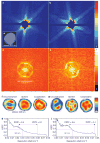
Figure 2. Single-shot diffraction patterns on single virus particles give interpretable results
a, b, Experimentally recorded far-field diffraction patterns (in false-colour representation) from individual virus particles captured in two different orientations. c, Transmission electron micrograph of an unstained Mimivirus particle, showing pseudo-icosahedral appearance. d, e, Autocorrelation functions for a (d) and b (e). The shape and size of each autocorrelation correspond to those of a single virus particle after high-pass filtering due to missing low-resolution data. f, g, Reconstructed images after iterative phase retrieval with the Hawk software package. The size of a pixel corresponds to 9 nm in the images. Three different reconstructions are shown for each virus particle: an averaged reconstruction with unconstrained Fourier modes and two averaged images after fitting unconstrained low-resolution modes to a spherical or an icosahedral profile, respectively. The orientation of the icosahedron was determined from the diffraction data. The results show small differences between the spherical and icosahedral fits. h, i, The PRTF for reconstructions where the unconstrained low-resolution modes were fitted to an icosahedron. All reconstructions gave similar resolutions. We characterize resolution by the point where the PRTF drops to 1/e (ref. 20). This corresponds to 32-nm full-period resolution in both exposures. Arrows mark the resolution range with other cut-off criteria found in the literature (Methods). Resolution can be substantially extended for samples available in multiple identical copies,–.

Comment in

  • Diffraction before destruction.
    Doerr A. Doerr A. Nat Methods. 2011 Apr;8(4):283. doi: 10.1038/nmeth0411-283. Nat Methods. 2011. PMID: 21574275 No abstract available.

References

    1. Neutze R, et al. Potential for biomolecular imaging with femtosecond X-ray pulses. Nature. 2000;406:752–757. - PubMed
    1. Chapman HN, et al. Femtosecond diffractive imaging with a soft-X-ray free-electron laser. Nature Phys. 2006;2:839–843.
    1. Bergh M, et al. Feasibility of imaging living cells at sub-nanometer resolution by ultrafast X-ray diffraction. Q Rev Biophys. 2008;41:181–204. - PubMed
    1. Nagler B, et al. Turning solid aluminium transparent by intense soft X-ray photoionization. Nature Phys. 2009;5:693–696.
    1. Emma P, et al. First lasing and operation of an ångstrom-wavelength free-electron laser. Nature Photon. 2010;4:641–647.

Publication types