. 2011 Mar;29(3):279-86.
doi: 10.1038/nbt.1783. Epub 2011 Feb 3.

A functionally characterized test set of human induced pluripotent stem cells

Affiliations

A functionally characterized test set of human induced pluripotent stem cells

Gabriella L Boulting et al. Nat Biotechnol. 2011 Mar.

Abstract

Human induced pluripotent stem cells (iPSCs) present exciting opportunities for studying development and for in vitro disease modeling. However, reported variability in the behavior of iPSCs has called their utility into question. We established a test set of 16 iPSC lines from seven individuals of varying age, sex and health status, and extensively characterized the lines with respect to pluripotency and the ability to terminally differentiate. Under standardized procedures in two independent laboratories, 13 of the iPSC lines gave rise to functional motor neurons with a range of efficiencies similar to that of human embryonic stem cells (ESCs). Although three iPSC lines were resistant to neural differentiation, early neuralization rescued their performance. Therefore, all 16 iPSC lines passed a stringent test of differentiation capacity despite variations in karyotype and in the expression of early pluripotency markers and transgenes. This iPSC and ESC test set is a robust resource for those interested in the basic biology of stem cells and their applications.


Figures
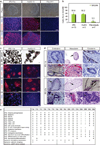
Figure 1
Figure 1. Characterization of pluripotency in the test-set of iPSC lines
(a) iPSC colonies were morphologically identical to hESC colonies, and express pluripotency markers NANOG and TRA-1-60, unlike the patient fibroblasts from which they were derived. Scale bars are 200 µm. (b) iPSC lines showed cell cycle profiles similar to that of ESCs and different to their parental fibroblasts. The percentage of cells undergoing different stages of the cell cycle was determined by propidium iodide staining and FC. The percentage of cells in S,G2 and M phase was found for each cell lines assayed, and then averaged for each category (c) Like hESCs, iPSC lines generated cell types of all three embryonic germ layers (endoderm- AFP, mesoderm- α-SMA, ectoderm- TUJ1) in vitro, as embryoid bodies (EBs), scale =100µm, and (d) when injected into mouse kidney capsules and allowed to form teratomas in vivo, scale=50µm. Representative images of H&E-stained sections are shown for lines 11b and 27e. Glands and goblet cells (endoderm), cartilage and muscle (mesoderm), pigmented neural epithelium and neural rosettes (ectoderm) are shown in the top and bottom panels respectively for both lines. (e) Summary chart depicting assays by which iPSC lines in the test set were characterized. Pluripotency assays (small +) for 29A and B were previously published by Dimos et al., 2008.
Figure 2
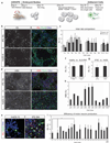
Figure 2. iPSCs show similar capacity for directed motor neuron differentiation to ESCs
(a) Protocol for directed differentiation of human stem cell lines into motor neurons. Cells were differentiated as EBs from day 0–29 in media formulations containing morphogens, including retinoic acid (RA), a small molecule agonist of the sonic hedgehog pathway (HAg), and neurotrophic factors BDNF, GDNF, and CNTF. EBs were dissociated and single cells plated for adherent culture on day 29. On day 32 cultures were analyzed. (b) Representative immunostaining results for iPSC (18c) and ESC (HuES-6) cultures show many ISL+ TUJ1+ motor neurons (scale = 50 µm). (c) The percentage of all nuclei which were ISL+ was quantified from differentiations performed independently in the Eggan and PALS labs. Data sets from lines differentiated in both labs are compared here, are highly similar, and have reproducible, characteristic %ISL efficiencies. 29e and 27e did not differentiate efficiently in either lab. (d) Efficiency of motor neuron differentiation was also measured by an alternative marker of motor neuron identity, HB9 (scale = 50 µm). (e) Many ISL+ motor neurons were also ChAT+, indicating proper maturation toward a cholinergic transmitter phenotype. (f) iPSC lines from control and ALS patients differentiated into ISL+ motor neurons with similar efficiencies, (g) as did ESCs and iPSCs. (h) The percentages of HB9+ nuclei were compared for a subset of iPSC lines and HuES-13. While comparisons again suggest donor- or line-specific differences, iPSC lines were overall equally capable of generating HB9+ motor neurons as HuES-13 (mean ± SD). (i) % ISL+ data from both labs was pooled for each iPSC and ESC line, and comparisons between lines showed generally similar performance, with significant differences between iPSC line 18c and iPSC lines 11a and 11c (p<0.05).
Figure 3
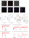
Figure 3. ESC and iPSC derived neurons are physiologically active
(a) Image of iPSC 11a-derived neurons filled with Fura Red AM and Fluo-4 AM dyes. The Fura Red channel is shown. The field illustrated is that imaged in panels b–g. Activity of labeled cells is represented in panels h and i. scale =100 µM. (b) ISL immunostaining of 11a field in a–g showing ISL+ neurons (star) and ISL- neurons (arrow). (c) Spontaneous electrical activity in cultured iPSC-derived neurons visualized by a ‘subtracted image’ that shows the difference in pixel intensities between two images acquired 1.7s apart in the Fluo 4 channel. Higher grey values represent increased pixel intensity. (d–g) Identically exposed pseudocolored averages of ten Fluo 4 AM images taken: (d) during the control period before addition of KA, (e) following treatment with 100 µM KA, (f) after washing following KA administration, and (g) following treatment with 50 mM KCl. Warmer colors represent increased fluorescence intensity. (h) Plot of Fluo 4/Fura Red intensity ratio in the somata of the two cells indicated by the star and arrow in a–c showing spontaneous activity. (i) Fluo 4/Fura Red intensity ratio of cells in a–c during sequential administration of KA and KCl indicated by bars above graph. (j) Examples of Fluo 4/Fura Red ratios from cell bodies of single spontaneously active cells in cultures of ESC R1-derived neurons, and iPSC 11a, 18a, 18c, and 27b-derived neurons as well as one example of a non-responsive, non-active cell in an R1 culture (NR). (k) Response of cells in (j) to KA and KCl. Sample voltage-clamp traces from (l) ESC and (m) iPSC 18a-derived neurons. (n) Blow-up of an iPSC 27b-derived neuron recording reveals typical sodium currents (left), which are blocked by 500 nM TTX (right). (o) Current-clamp recordings of single action potentials in ESC and iPSC 27b-derived neurons as well as multiple action potentials in an iPSC 18a-derived neuron.
Figure 4
Figure 4. Persistent transgene expression does not inhibit differentiation
(a) Quantitative RT-PCR was used to measure relative levels of transcript from endogenous genes ‘e’ and viral transgenes ‘v’ of the reprogramming factors OCT4 and KLF4 in undifferentiated iPSCs and ESCs, and in day 32 neuron cultures. Transgene expression or silencing in the undifferentiated cells is maintained after differentiation. Relative levels in undifferentiated HuES-3 were set as 1. (b) Day 32 motor neuron cultures were co-stained for ISL and OCT4. HuES-3 and iPSC 17a-derived cultures, which do not express viral OCT4, did not stain for OCT4. However, iPSC 15b-derived cultures, which do express viral OCT4, contained many OCT4+ ISL+ motor neurons and OCT4+ISL+ cells. Arrow = OCT4+ ISL+, arrow head = OCT4+ ISL, chevron = OCT4 ISL+.
Figure 5
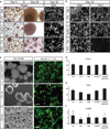
Figure 5. Sub-optimal iPSC lines can be rescued using SMAD inhibition
(a) During EB differentiation, iPSC lines 27e and 29e showed abnormal EB morphology and survival compared to lines that behaved normally (HuES-3 and 29d shown), phase scale = 500 µm, DNA scale = 129 µm. (b) Although EBs from iPSC line 11b had typical morphology, day 32 cultures showed decreased neuronal TUJ1 staining compared to all other normal lines (HuES-13 and 18a shown), scale = 129 µm. (c) Representative phase and immunostaining images for previously defective iPSC lines 29e, 11b, and control ESC lines HuES-3 and HuES-3-hb9:GFP. Phase image scales are 500 µm, immunostaining image scales are 100 µm. Quantification of immunostaining in differentiated cultures derived from the three previously problematic iPSC lines (11b, 27e, 29e) and ESC controls (d) percentage of TUJ1+ cells; (e) percentage of ISL+ cells; (f) percentage of HB9+ cells. Mean ± SEM.

Comment in

  • IPSCs put to the test.
    Kim H, Studer L. Kim H, et al. Nat Biotechnol. 2011 Mar;29(3):233-5. doi: 10.1038/nbt.1805. Nat Biotechnol. 2011. PMID: 21390027 No abstract available.
  • Testing pluripotency.
    de Souza N. de Souza N. Nat Methods. 2011 Apr;8(4):287. doi: 10.1038/nmeth0411-287. Nat Methods. 2011. PMID: 21574272 No abstract available.

Similar articles

Cited by

References

    1. Kiskinis E, Eggan K. Progress toward the clinical application of patient-specific pluripotent stem cells. J Clin Invest. 2010;120:51–59. - PMC - PubMed
    1. Dimos JT, et al. Induced pluripotent stem cells generated from patients with ALS can be differentiated into motor neurons. Science. 2008;321:1218–1221. - PubMed
    1. Park IH, et al. Disease-specific induced pluripotent stem cells. Cell. 2008;134:877–886. d. - PMC - PubMed
    1. Lee G, et al. Modelling pathogenesis and treatment of familial dysautonomia using patient-specific iPSCs. Nature. 2009;461:402–406. - PMC - PubMed
    1. Soldner F, et al. Parkinson's disease patient-derived induced pluripotent stem cells free of viral reprogramming factors. Cell. 2009;136:964–977. - PMC - PubMed

Publication types