

Enhanced NMDA receptor NR1 phosphorylation and neuronal activity in the arcuate nucleus of hypothalamus following peripheral inflammation
- PMID: 21293467
- PMCID: PMC4009944
- DOI: 10.1038/aps.2010.190
Enhanced NMDA receptor NR1 phosphorylation and neuronal activity in the arcuate nucleus of hypothalamus following peripheral inflammation
Abstract
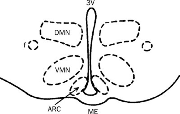
AbstractAim:To investigate the role of glutamate and N-methyl-D-aspartate (NMDA) receptors in central sensitization following peripheral inflammation in the arcuate nucleus (ARC) of the mediobasal hypothalamus.
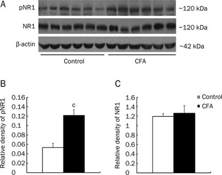
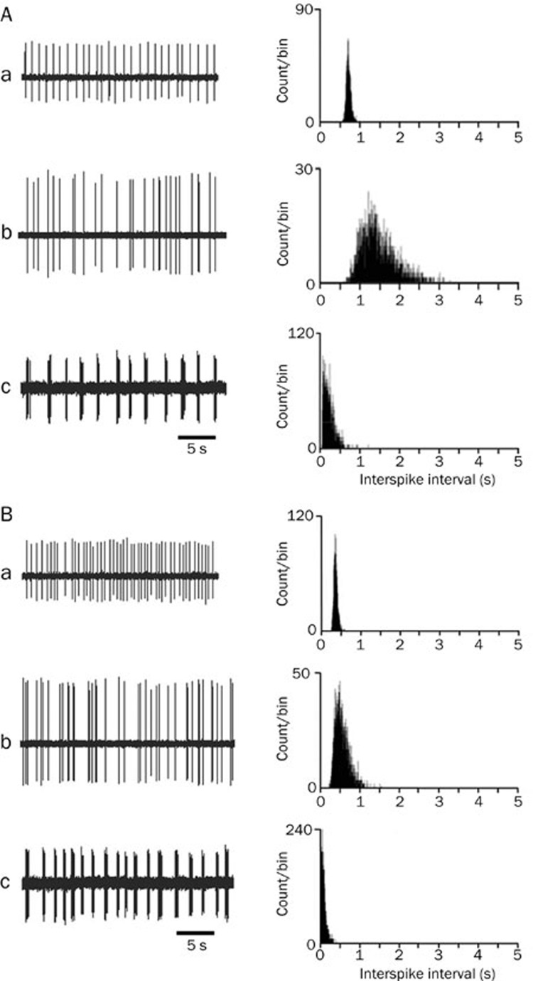
Methods: Mediobasal hypothalamic slices were prepared from rats undergoing peripheral inflammation, which was induced by a unilateral injection of complete Freund's adjuvant (CFA) into hind paw. Neuronal activation levels in the ARC were monitored by recording extracellular unit discharges. The NMDA receptor NR1 subunit (NR1) was measured using Western blot analysis.
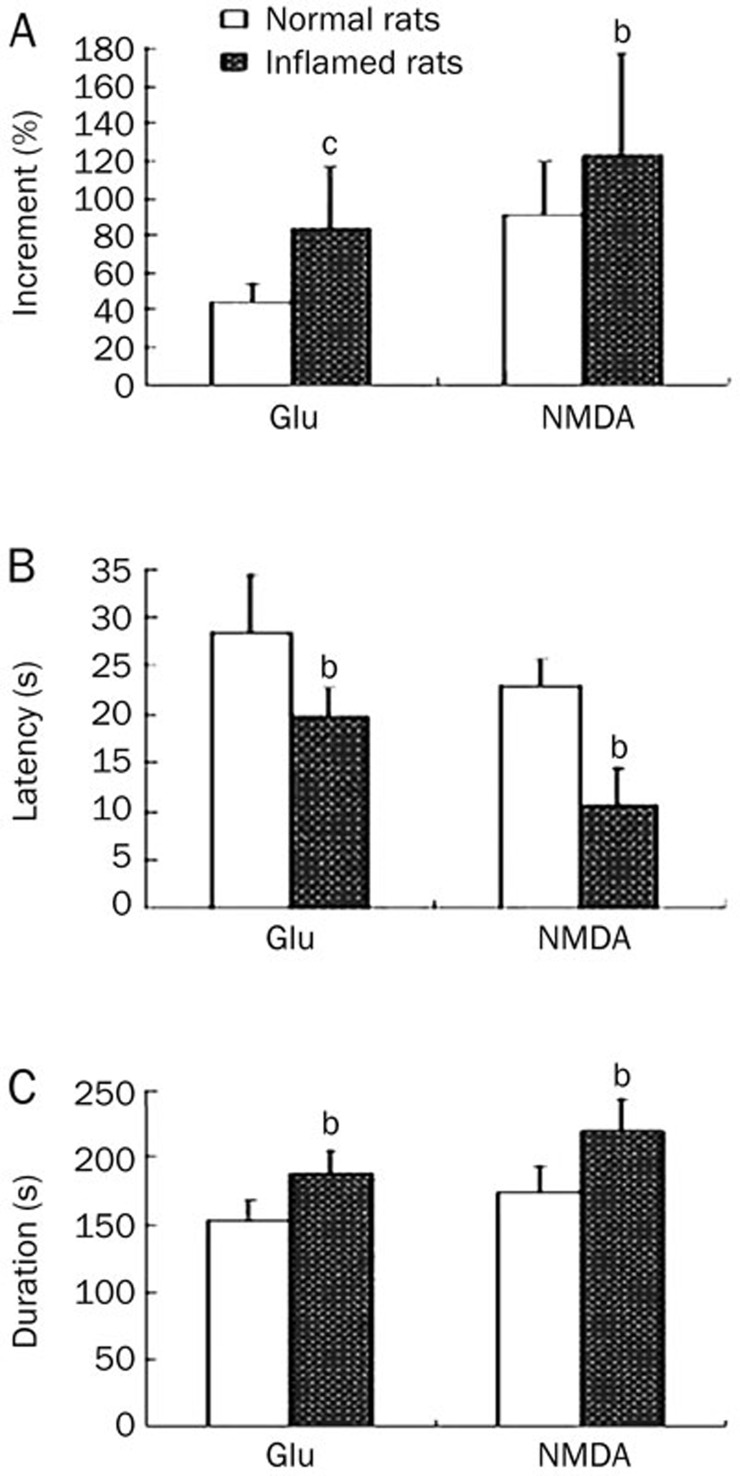
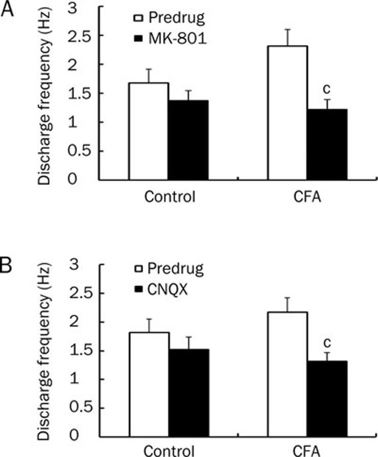
Results: Enhanced NR1 phosphorylation was observed in the ARC of CFA-inflamed rats. Compared with the control rats, the firing rate of spontaneous discharges in ARC neurons of inflamed rats was significantly higher, and it was significantly reduced both by an NMDA receptor antagonist (MK-801, 300 μmol/L) and by a non-NMDA receptor antagonist (CNQX, 30 μmol/L). Application of exogenous glutamate (200 μmol/L) or NMDA (25 μmol/L) resulted in increased neuronal discharges for ARC neurons, which was enhanced to a greater extent in inflamed rats than in control rats.
Conclusion: Glutamate receptor activation in the hypothalamic ARC plays a crucial role in central sensitization associated with peripheral inflammation.
Figures
References
-
- Dingledine K, Borges K, Bowie D, Traynelis S. The glutamate receptor ion channels. Pharmacol Rev. 1999;51:7–61. - PubMed
-
- Fundytus ME. Glutamate receptors and nociception: implications for the drug treatment of pain. CNS Drugs. 2001;15:29–58. - PubMed
-
- Petrenko AB, Yamakura T, Baba H, Shimoji K. The role of N-methyl-D-aspartate (NMDA) receptors in pain: a review. Anesth Analg. 2003;97:1108–16. - PubMed
-
- Rygh LJ, Svendsen F, Hole K, Tjolsen A. Natural noxious stimulation can induce long-term increase of spinal nociceptive responses. Pain. 1999;82:305–10. - PubMed
-
- Sandkuhler J, Liu X. Induction of long-term potentiation at spinal synapse by noxious stimulation or nerve injury. Eur J Neurosci. 1998;10:2476–80. - PubMed
Publication types
MeSH terms
Substances
LinkOut - more resources
Full Text Sources
