

Comparison of retinal thickness values and segmentation performance of different OCT devices in acute branch retinal vein occlusion
- PMID: 21293498
- PMCID: PMC3171252
- DOI: 10.1038/eye.2010.216
Comparison of retinal thickness values and segmentation performance of different OCT devices in acute branch retinal vein occlusion
Abstract
Purpose: To compare retinal thickness (RT) measurement and segmentation performance of time domain (TD, Stratus) and spectral domain (SD) optical coherence tomography (OCT) devices (Cirrus, Spectralis) for imaging macular oedema (ME) secondary to branch retinal vein occlusion (BRVO).
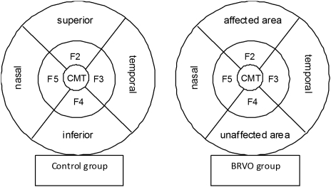
Methods: In this study, 20 eyes of 20 consecutive patients with acute BRVO were included. A total of 18 unaffected fellow eyes served as control group. RT measurement was analysed in the five inner fields of the early-treatment diabetic retinopathy grid, and proportional segmentation errors were evaluated.
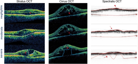
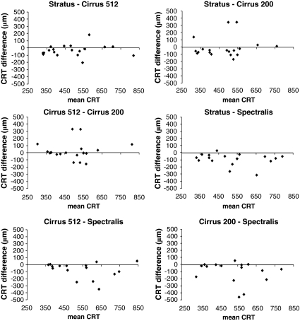
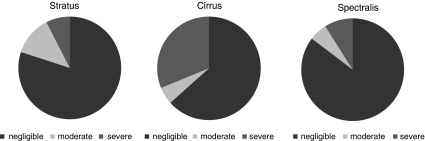
Results: Central millimetre thickness (CMT) showed a mean difference of -64, -74, and -18 μm (P < 0.001) in the control group and -31 μm (P=0.107), -92 μm (P<0.001), and -105 μm (P=0.016) in the BRVO group, between Stratus and Cirrus, between Stratus and Spectralis, and between Cirrus and Spectralis, respectively. Mean RT showed the highest variability between different devices in the area most intensively affected by BRVO-related ME. In eyes with BRVO, 14.6% of Spectralis, 20% of Stratus, and 36.6% of Cirrus scans demonstrated moderate and severe segmentation errors.
Conclusion: RT measurement in eyes with BRVO, by TD and SD OCT, is compromised by a significant rate of segmentation errors. Deviations are most pronounced in the areas most severely affected by ME.
Figures
 ) segmentation errors.
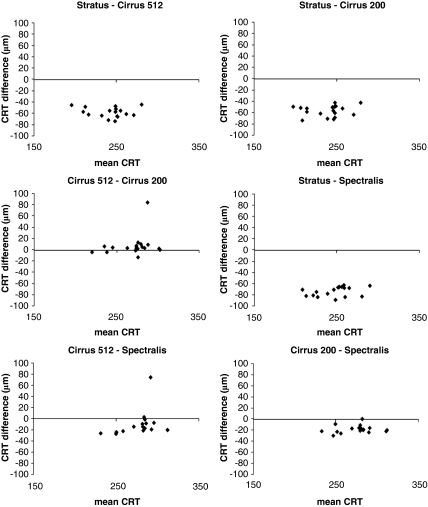
References
-
- Mitchell P, Smith W, Chang A. Prevalence and associations of retinal vein occlusion in Australia. The Blue Mountains Eye Study. Arch Ophthalmol. 1996;114:1243–1247. - PubMed
-
- Yamaike N, Tsujikawa A, Ota M, Sakamoto A, Kotera Y, Kita M, et al. Three-dimensional imaging of cystoid macular edema in retinal vein occlusion. Ophthalmology. 2008;115:355–362. - PubMed
-
- Prager F, Michels S, Kriechbaum K, Georgopoulos M, Funk M, Geitzenauer W, et al. Intravitreal bevacizumab (Avastin) for macular edema secondary to retinal vein occlusion: 12-month results of a prospective clinical trial. Br J Ophthalmol. 2009;93:452–456. - PubMed
MeSH terms
LinkOut - more resources
Full Text Sources
