Case Reports
. 2011 Jan 29:17:309-22.

Phenotypic variability in a French family with a novel mutation in the BEST1 gene causing multifocal best vitelliform macular dystrophy

Affiliations
Case Reports

Phenotypic variability in a French family with a novel mutation in the BEST1 gene causing multifocal best vitelliform macular dystrophy

Emmanuelle Lacassagne et al. Mol Vis. .

Abstract

Aims: To describe genetic and clinical findings in a French family affected by best vitelliform macular dystrophy (BVMD).

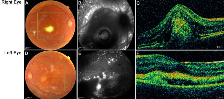
Methods: We screened eight at-risk members of a family, including a BVMD-affected proband, by direct sequencing of 11 bestrophin-1 (BEST1) exons. Individuals underwent ophthalmic examination and autofluorescent fundus imaging, indocyanine green angiography, electro-oculogram (EOG), electroretinogram (ERG), multifocal ERG, optical coherence tomography (OCT), and where possible, spectral domain OCT.

Results: The sequence analysis of the BEST1 gene revealed one previously unknown mutation, c.15C>A (p.Y5X), in two family members and one recently described mutation, c.430A>G (p.S144G), in five family members. Fundus examination and electrophysiological responses provided no evidence of the disease in the patient carrying only the p.Y5X mutation. Three patients with the p.S144G mutation did not show any preclinical sign of BVMD except altered EOGs. Two individuals of the family exhibited a particularly severe phenotype of multifocal BVMD-one individual carrying the p.S144G mutation heterozygously and one individual harboring both BEST1 mutations (p.S144G inherited from his mother and p.Y5X from his father). Both of these family members had multifocal vitelliform autofluorescent lesions combined with abnormal EOG, and the spectral domain OCT displayed a serous retinal detachment. In addition, ERGs demonstrated widespread retinal degeneration and multifocal ERGs showed a reduction in the central retina function, which could be correlated with the decreased visual acuity and visual field scotomas.

Conclusions: A thorough clinical evaluation found no pathological phenotype in the patient carrying the isolated p.Y5X mutation. The patients carrying the p.S144G variation in the protein exhibited considerable intrafamilial phenotypic variability. Two young affected patients in this family exhibited an early onset, severe, multifocal BVMD with a diffuse distribution of autofluorescent deposits throughout the retina and rapid evolution toward the loss of central vision. The other genetically affected relatives had only abnormal EOGs and displayed no or extremely slow electrophysiological evolution.


Figures

Figure 1
Figure 1
Pedigree of the family studied and segregation of the BEST1 mutant alleles. This figure shows the pedigree of a french family displaying an unusual phenotype reminiscent of very atypic bestrophinopathy. The clinical status of I-1 and I-2 are unknown. The red shapes denote genotyped individuals. White circles represent unaffected females, filled circles affected females, white squares represent unaffected males and filled squares affected males. The p.Y5X mutation is shown in gray and the p.S144G mutation is shown in black. Individual III-1 (the proband) harbors both mutations.
Figure 2
Figure 2
Two novel nucleotidic mutations in the BEST1 gene. Electrophoregrams of the BEST1 gene mutations found in the affected members of the French family studied and phylogenetic conservation throughout evolution of the normal BEST-1 amino-acid residues affected by these mutations. A: These electrophoregrams show heterozygous mutated nucleotides in the BEST1 gene: An adenine (A) is replaced by a guanine (G) at the 430th nucleotidic position of the BEST1 cDNA sequence (c.430A>G) and and a cytosine (C) is replaced by an adenine (A) at the 15th nucleotidic position of the BEST1 cDNA sequence (c.15C>A) (top panel), and normal sequences (low panel). The peaks in red indicate thymidine (T), green indicate A, black indicate G, and blue indicate C. B: This panel shows the multiple sequence alignment of human bestrophin-1 protein (BEST-1 protein; NP_004174) with the BEST-1 protein sequences from Mus musculus (NP_036043.2), Rattus norvegicus (NP_001011940.1), Xenopus tropicalis (BAH70274.1), and Drosophila melanogaster (AAF54503.1). This multiple sequence alignment highlights the strong conservation throughout evolution of the amino-acid residues of the normal BEST-1 protein which were found affected by mutations in this study. C: This panel shows the multiple sequence alignment of the human BEST1 protein with the bestrophin paralogs: BEST2, BEST3, and BEST4. Alignments are zoomed into the relevant region. The amino- acids affected by a mutation are shown in red. The stars indicate 100% conservation.
Figure 3
Figure 3
Color fundus and autofluorescent fundus/optical coherence tomography scans in patients II-1, II-2, II-3, III-2, and III-3. No macular lesion was detectable by autofluorescence imaging or optical coherence tomography scanning for patients II-1 or III-2. No macular lesion was detectable in the color fundus for patients II-2, II-3, or III-3.
Figure 4
Figure 4
Right and left eye color fundus, autofluorescent fundus, indocyanine green angiography, and optical coherence tomography scans in the proband (patient III-1). Well demarcated vitelliform lesions in the central macula are detected by fundoscopy (A-E) and are also apparent on the autofluorescence image (B-F) and indocyanine green angiography (C-G) in both eyes. Optical coherence tomography images through the fovea show a highly reflective thickened layer at the level of the retinal pigment epithelium and choriocapillaris of both eyes and well circumscribed elevation of the retinal pigment epithelium in both eyes (D-H).
Figure 5
Figure 5
Fundoscopy, autofluorescence and optical coherence tomography (OCT3) imaging of a severely affected patient (III-4). Typical vitelliform lesions are visible on the ophthalmoscopic appearance of the right eye (A, B, C) and left eye shows fragmented vitelliform lesions (D, E, F). Green lines indicate abrupt transitions and the frame of the fundus that was scanned by optical coherent tomography (OCT). The middle green lines of (A, D) indicate the horizontal axis of the OCT scan shown in (C, F).
Figure 6
Figure 6
Electrophysiology measurements of three representative cases of the family studied. This figure represents electro-oculograms (EOGs; A), right eye flash electroretinograms (ERGs; B) and right eye multifocal electroretinograms (mfERGs; C) in patients carrying one mutation heterozygously (II-1: p.Y5X; III-2: p.S144G) or both mutations (III-1). Findings are based on ISCEV standard. Patients II-2 and II-3 displayed electrophysiological findings similar to III-2 and patient III-4 displayed electrophysiological findings similar to III-1. Except for II-1, the amplitudes for the light phase of the EOG (A) were abnormal with a reduction in the Arden ratio (EOG light rise <150%). In patient III-1 (and III-4), flash ERGs show generalized decreased rod and cone photoreceptor amplitudes and decreased photopic oscillatory potentials amplitude (Phot-Ops; B). MfERG records a reduced central function with relative preservation of the amplitude response and timing from the surrounding macula (C).

Similar articles

Cited by

References

    1. Marmorstein AD, Cross HE, Peachey NS. Functional roles of bestrophins in ocular epithelia. Prog Retin Eye Res. 2009;28:206–26. - PMC - PubMed
    1. Petrukhin K, Koisti MJ, Bakall B, Li W, Xie G, Marknell T, Sandgren O, Forsman K, Holmgren G, Andreasson S, Vujic M, Bergen AA, McGarty-Dugan V, Figueroa D, Austin CP, Metzker ML, Caskey CT, Wadelius C. Identification of the gene responsible for Best macular dystrophy. Nat Genet. 1998;19:241–7. - PubMed
    1. Marquardt A, Stohr H, Passmore LA, Kramer F, Rivera A, Weber BH. Mutations in a novel gene, VMD2, encoding a protein of unknown properties cause juvenile-onset vitelliform macular dystrophy (Best's disease). Hum Mol Genet. 1998;7:1517–25. - PubMed
    1. Krämer F, White K, Pauleikhoff D, Gehrig A, Passmore L, Rivera A, Rudolph G, Kellner U, Andrassi M, Lorenz B, Rohrschneider K, Blankenagel A, Jurklies B, Schilling H, Schütt F, Holz FG, Weber BH. Mutations in the VMD2 gene are associated with juvenile-onset vitelliform macular dystrophy (Best disease) and adult vitelliform macular dystrophy but not age-related macular degeneration. Eur J Hum Genet. 2000;8:286–92. - PubMed
    1. Lotery AJ, Munier FL, Fishman GA, Weleber RG, Jacobson SG, Affatigato LM, Nichols BE, Schorderet DF, Sheffield VC, Stone EM. Allelic variation in the VMD2 gene in best disease and age-related macular degeneration. Invest Ophthalmol Vis Sci. 2000;41:1291–6. - PubMed

Publication types

MeSH terms

LinkOut - more resources