. 2011 Nov;24(9):1111-8.
doi: 10.1002/nbm.1666. Epub 2011 Feb 4.

Background suppression in arterial spin labeling MRI with a separate neck labeling coil

Affiliations

Background suppression in arterial spin labeling MRI with a separate neck labeling coil

Qiang Shen et al. NMR Biomed. 2011 Nov.

Abstract

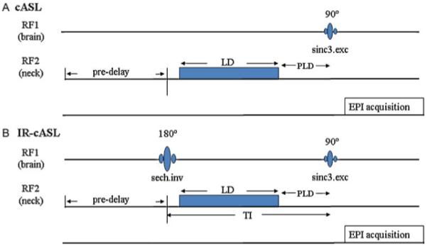
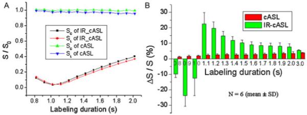
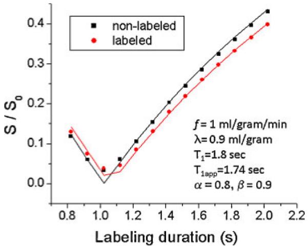
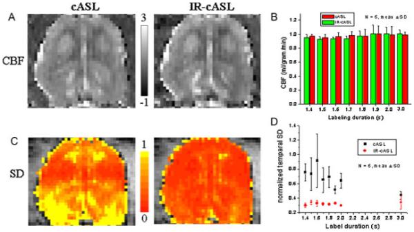
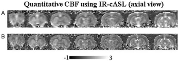
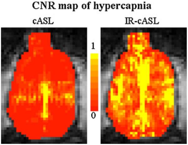
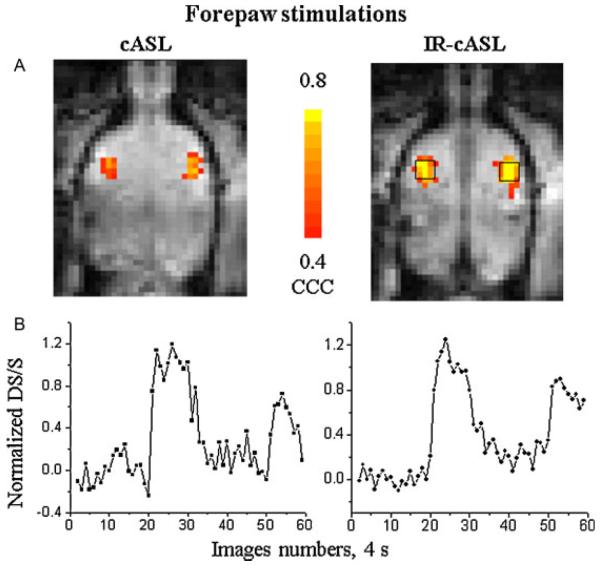
In arterial spin labeling (ASL) MRI to measure cerebral blood flow (CBF), pair-wise subtraction of temporally adjacent non-labeled and labeled images often can not completely cancel the background static tissue signal because of temporally fluctuating physiological noise. While background suppression (BS) by inversion nulling improves CBF temporal stability, imperfect pulses compromise CBF contrast. Conventional BS techniques may not be applicable in small animals because the arterial transit time is short. This study presents a novel approach of BS to overcome these drawbacks using a separate 'neck' radiofrequency coil for ASL and a 'brain' radiofrequency coil for BS with the inversion pulse placed before spin labeling. The use of a separate 'neck' coil for ASL should also improve ASL contrast. This approach is referred to as the inversion-recovery BS with the two-coil continuous ASL (IR-cASL) technique. The temporal and spatial contrast-to-noise characteristics of basal CBF and CBF-based fMRI of hypercapnia and forepaw stimulation in rats at 7 Tesla were analyzed. IR-cASL yielded two times better temporal stability and 2.0-2.3 times higher functional contrast-to-noise ratios for hypercapnia and forepaw stimulation compared with cASL without BS in the same animals. The Bloch equations were modified to provide accurate CBF quantification at different levels of BS and for multislice acquisition where different slices have different degree of BS and residual degree of labeling. Improved basal CBF and CBF-based fMRI sensitivity should lead to more accurate CBF quantification and should prove useful for imaging low CBF conditions such as in white matter and stroke.


Figures

Figure 1
Figure 1
Pulse sequence diagram for (A) continuous arterial spin labeling (cASL) and (B) inversion recovery (IR)-cASL. Inversion pulse for the background suppression is transmitted via the brain coil on the first radiofrequency (RF) channel. The ASL is transmitted via a separate neck coil on the second RF channel. The inversion delay TI is the sum of labeling duration (LD) and post-labeling delay (PLD), half the inversion pulse length and half of the excitation pulse length. The pre-delay is used for magnetization to return to equilibrium. Image acquisition uses gradient-echo echo-planar imaging.
Figure 2
Figure 2
(A) Normalized Snon-labeled and Slabeled signal intensities as a function of labeling durations for inversion recovery continuous arterial spin labeling (IR-cASL) and cASL acquisition from one animal. Normalization was taken with respective to the mean value of Snon-labeled of cASL scans. (B) Group-averaged ΔS/S in percentage as a function of labeling durations of the cASL and IR-cASL acquisition. Normalization was taken with respective to each method’s non-labeled signal.
Figure 3
Figure 3
Modeling of eqns [5] and [10] on experimental data. Snon-labeled and Slabeled signal intensities are plotted as a function of labeling duration of the inversion recovery continuous arterial spin labeling (IR-cASL) experiments. Data points were experimental data. Lines were derived from eqns [5] and [10] using parameters as shown.
Figure 4
Figure 4
(A) Cerebral blood flow (CBF) images of the continuous arterial spin labeling (cASL) and inversion recovery (IR)-cASL acquisition (horizontal view). (B) CBF values versus labeling durations. Scale bars indicate CBF units in mL/g/min. (C) Temporal standard deviation (SD) maps of cASL and IR-cASL acquisition. (D) Temporal SDs of cASL and IR-cASL versus labeling durations.
Figure 5
Figure 5
Multislice cerebral blood flow (CBF) images (ml/g/min) by inversion recovery continuous arterial spin labeling (IR-cASL) at (A) 490×490 μm and (B) 245×245 μm (transverse view). The total acquisition time was 3 mins for each resolution.
Figure 6
Figure 6
Contrast-to-noise ratio (CNR) maps associated with hypercapnic challenges of the continuous arterial spin labeling (cASL) and inversion recovery (IR)-cASL acquisition (horizontal view).
Figure 7
Figure 7
(A) Cerebral blood flow (CBF) fMRI activation maps and (B) time courses associated with forepaw stimulations of the continuous arterial spin labeling (cASL) and inversion recovery (IR)-cASL acquisition (horizontal view). These maps were obtained using identical statistical threshold. Time courses were obtained from the same region of interest for both cASL and IR-cASL as shown.

References

    1. Roy CS, Sherrington CS. On the regulation of blood supply of the brain. J. Physiol. 1890;1:85–108. - PMC - PubMed
    1. Williams DS, Detre JA, Leigh JS, Korestsky AP. Magnetic resonance imaging of perfusion using spin inversion of arterial water. Proc. Natl. Acad. Sci. USA. 1992;89:212–216. - PMC - PubMed
    1. Barbier EL, Lamalle L, Decorps M. Methodology of brain perfusion imaging. J. Magn. Reson. Imaging. 2001;13:496–520. - PubMed
    1. Duong TQ, Kim D-S, Ugurbil K, Kim S-G. Localized blood flow response at sub-millimeter columnar resolution. Proc. Natl. Acad. Sci. USA. 2001;98:10904–10909. - PMC - PubMed
    1. Dixon WT, Sardashti M, Castillo M, Stomp GP. Multiple inversion recovery reduces static tissue signal in angiograms. Magn. Reson. Med. 1991;18:257–268. - PubMed

Publication types