. 2011 Mar 2;133(8):2583-91.
doi: 10.1021/ja108846h. Epub 2011 Feb 4.

Deep penetration of a PDT drug into tumors by noncovalent drug-gold nanoparticle conjugates

Affiliations

Deep penetration of a PDT drug into tumors by noncovalent drug-gold nanoparticle conjugates

Yu Cheng et al. J Am Chem Soc. .

Abstract

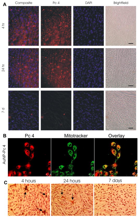
Efficient drug delivery to tumors is of ever-increasing importance. Single-visit diagnosis and treatment sessions are the goal of future theranostics. In this work, a noncovalent PDT cancer drug-gold nanoparticle (Au NP) conjugate system performed a rapid drug release and deep penetration of the drug into tumors within hours. The drug delivery mechanism of the PDT drug through Au NPs into tumors by passive accumulation was investigated via fluorescence imaging, elemental analysis, and histological staining. The pharmacokinetics of the conjugates over a 7-day test period showed rapid drug excretion, as monitored via the fluorescence of the drug in urine. Moreover, the biodistribution of Au NPs in this study period indicated clearance of the NPs from the mice. This study suggests that noncovalent delivery via Au NPs provides an attractive approach for cancer drugs to penetrate deep into the center of tumors.


Figures

Figure 1
Figure 1
A) Schematic of Au NP-Pc 4 conjugates circulation into the tumor. After circulating within the blood-vascular system, Au NP-Pc 4 extravasate through the endothelial cell layer of the blood vessels into cancerous tissues via the EPR effect. B) Chemical structure of the PDT drug Pc 4. C) The hydrodynamic diameter of the Au NP-Pc 4 conjugates measured by dynamic light scattering (DLS).
Figure 2
Figure 2
In vivo fluorescence imaging of Au NP-Pc 4 conjugates. Tumor bearing mice were injected intravenously with Au NP-Pc 4 conjugates at a Pc 4 dosage of 1 mg kg−1 mouse. (A) In vivo fluorescence imaging of the Au NP-Pc 4 conjugates injected mouse at various time points within 24 hours. Arrows indicate tumor location. (B) The average fluorescence intensities from the tumor areas of 24-hour post-injection mice (n=5). (C) Picture of the tumor at 4 hours post-injection (left) and the corresponding 3D surface plot (right) of pixel intensities (Pc 4 fluorescence) obtained from ImageJ. (D) Comparison of the fluorescence images in whole versus transected tumors at 4 hours, 24 hours and 7 days post-injection.
Figure 3
Figure 3
Au NP-Pc 4 accumulation in subcutaneous heterotopic tumors. (A) Cryosections of ex vivo tumors were counterstained with DAPI (blue) to visualize nuclei. Pc 4 fluorescence was captured using a standard Cy5 filter set (red). Images were overlaid to demonstrate Pc 4 uptake into the tumor. All images were captured at 40X magnification. Scale bar represents 100 μm. (B) Confocal images of the cancer cells incubated with Au NP-Pc 4 for 4 hours stained with mitotracker green. (C) Au NPs visualized using silver enhancement staining in ex vivo paraffin-embedded tumor sections 4 hours, 24 hours, and 7 days post-injection. Arrows indicate the blood vasculatures. The size of Au NPs was enhanced by deposition of the elemental silver on the NP surface.
Figure 4
Figure 4
Au NP-Pc 4 accumulation in subcutaneous heterotopic tumors. (A) Graph of average Pc 4 fluorescence intensity; inset graph of Au concentration in tumors from the injected mice over a 7 day period (n=3). (B) Gold concentration in blood based on total injected dose at different time points in Au NP-Pc 4 injected mice (n=5).
Figure 5
Figure 5
Biodistribution and elimination of Au NP-Pc 4 based on Pc 4 fluorescence. The Pc 4 fluorescence images of organs from the injected mice after dissection.
Figure 6
Figure 6
Assessment of Au NP-Pc 4 biodistribution on Au content. (A) Au (μg) per gram of sample in organs. Mice were injected with Au NP-Pc 4 (n≥3 per each time point). The total gold content in tissue samples was evaluated by GFAAS. (B) Histology studies of the organ tissues. Organ samples were removed from mice injected with Au NP-Pc 4 after 4 hours, 24 hours, and 7 days post-injection. The paraffin-embedded tissue slices were stained with hematoxylin, eosin, and silver enhancement reagents. The black and brownish spots indicate Au NPs in the tissues. Images were captured at 40X magnification. Scale bar represents 100 μm.

References

    1. Heldin CH, Rubin K, Pietras K, Ostman A. Nat Rev Cancer. 2004;4:806. - PubMed
    1. Sugahara KN, Teesalu T, Karmali PP, Kotamraju VR, Agemy L, Girard OM, Hanahan D, Mattrey RF, Ruoslahti E. Cancer Cell. 2009;16:510. - PMC - PubMed
    1. Langer R. Nature. 1998;392:5. - PubMed
    1. Torchilin VP. Adv Drug Deliv Rev. 2006;58:1532. - PubMed
    1. Peer D, Karp JM, Hong S, Farokhzad OC, Margalit R, Langer R. Nat Nanotechnol. 2007;2:751. - PubMed

Publication types

MeSH terms