. 2011 Oct;20(10):1649-58.
doi: 10.1089/scd.2010.0455. Epub 2011 Mar 12.

Isolation of live label-retaining cells and cells undergoing asymmetric cell division via nonrandom chromosomal cosegregation from human cancers

Affiliations

Isolation of live label-retaining cells and cells undergoing asymmetric cell division via nonrandom chromosomal cosegregation from human cancers

Danielle Hari et al. Stem Cells Dev. 2011 Oct.

Abstract

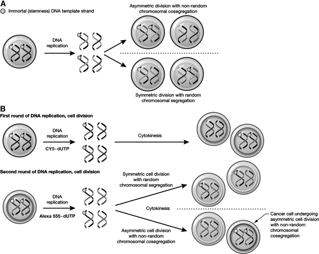
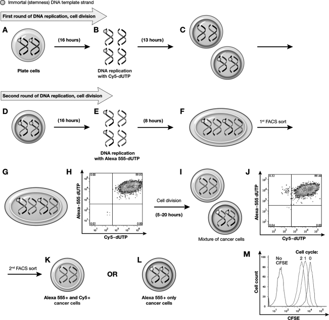
The ability to retain DNA labels over time is a property proposed to be associated with adult stem cells. Recently, label retaining cells (LRC) were indentified in cancer. LRC were suggested to be the result of either slow-cycling or asymmetric-cell-division with nonrandom-chromosomal-cosegregation (ACD-NRCC). ACD-NRCC is proposed to segregate the older template DNA strands into daughter stem cells and newly synthesized DNA into daughter cells destined for differentiation. The existence of cells undergoing ACD-NRCC and the stem-like nature of LRC remain controversial. Currently, to detect LRC and ACD-NRCC, cells need to undergo fixation. Therefore, testing the stem-cell nature and other functional traits of LRC and cells undergoing ACD-NRCC has been limited. Here, we show a method for labeling DNA with single and dual-color nucleotides in live human liver cancer cells avoiding the need for fixation. We describe a novel methodology for both the isolation of live LRC and cells undergoing ACD-NRCC via fluorescence-activated cell sorting with confocal microscopy validation. This has the potential to be a powerful adjunct to stem-cell and cancer research.


Figures

FIG. 1.
FIG. 1.
The immortal strand hypothesis and detection of ACD-NRCC by dual nucleotide-analogue DNA labeling. (A) The immortal strand hypothesis proposes that each chromosome in a stem-cell has 1 template-DNA strand that can be conserved after numerous asymmetric cell divisions (orange dot). These strands are preferentially cosegregated into daughter stem-cells by ACD-NRCC, whereas chromosomes that do not carry these strands segregate into daughter cells fated for differentiation. (B) To detect ACD-NRCC by dual nucleotide-analog DNA labeling procedure, cells are labeled sequentially for 2 cell cycles with 2 different nucleotides; first Cy5 (pseudo-colored green) then Alexa555 (red). When symmetric division occurs, both nuclei of the daughter cells will incorporate both nucleotides. However, when ACD-NRCC occurs, 1 nucleus will incorporate both Cy5 and Alexa555 (yellow), whereas the other nucleus will incorporate only Alexa555. ACD-NRCC, asymmetric cell division with nonrandom chromosomal cosegregation.
FIG. 2.
FIG. 2.
Isolation of live cells undergoing ACD-NRCC. After the schema described in Fig. 1B, for the first cell cycle of DNA replication, liver cancer cells were plated (A), stained with CFSE (B), and labeled with Cy5 (C). Subsequently, cells that underwent exactly 1 cell division (50% CFSE; D, E) and were Cy5+ High (F) were sorted by FACS with 99% purity (G, H). For the second round of DNA replication, cells were labeled with Alexa555 (I). After completion of the second cycle of DNA replication (J), cells that underwent exactly 2 cell divisions (25% CFSE) were sorted (K) and were Alexa555+ high and Cy5 high (L, M). Among these cells, 2 populations were identified and sorted: 25% CFSE+/Alexa555+ high cells (N, O) and 25% CFSE+/Alexa555+/Cy5+ high cells (P–R). The 25% CFSE+/Alexa555+ subpopulation represents cells that were generated by ACD-NRCC. The 25% CFSE+/Alexa555+/Cy5+ subpopulation represents the cells in which random chromosomal segregation has occurred (symmetric division). CFSE, carboxyfluorescein diacetate succinimidyl ester; FACS, fluorescence-activated cell sorting.
FIG. 3.
FIG. 3.
Isolation of live cells undergoing ACD-NRCC (second method). For the first cell cycle of DNA replication, liver cancer cells were plated (A), labeled with Cy5 (B), and were let to complete the first cell division in culture (C). Subsequently, cells completing the first round of DNA replication were labeled with Alexa555 (D,E). Before completion of the second round of DNA replication (F), cells that were Alexa555-high/Cy5-high and undergoing mitosis were sorted by FACS and placed in a culture to complete mitosis (G, H). At the completion of mitosis of the second round of DNA replication (I), cultured cells that underwent 2 cell divisions (25% CFSE) were analyzed and subsequently sorted (J). The cultured cells that previously contained only Alexa555-high/Cy5-high cells now generated 2 populations of cells: CFSE-25%/Alexa555-high/Cy5-high cells generated by symmetric division (K), and CFSE25%/Alexa555-high only cells generated by ACD-NRCC (L). CFSE was used to detect contemporaneously cell division status (M).
FIG. 4.
FIG. 4.
Three-dimensional fluorescent confocal microscopy capturing symmetric and asymmetric cell division in live cells. To confirm the results described in Fig. 2, we tested the products the cell sorting after each stage. (A) shows cells that were isolated by FACS after the first round of DNA replication, [50% CFSE+ (pseudo-colored white)/Cy5+ (pseudo-colored green) cells]. (B, C) shows the 2 populations of cells isolated after the second round of DNA replication: 25% CFSE+ (white)/Cy5+(green)/Alexa555+(red) cells (B) and cells that underwent ACD-NRCC 25% CFSE+(white)/Alexa555+(red) cells (C). (D) Live cells undergoing symmetric cell division containing 2 nuclei labeled with both nucleotides during mitosis (green and red). (E) Live cell undergoing ACD-NRCC is illustrated containing 1 nucleus with the second labeled nucleotides only (red) and the other nucleus containing both nucleotides (green and red). The DAPI (blue) reveals 2 nuclei in the same cytoplasmic space (white) without an intervening membrane. (F) Shows the same cell from E during mitosis where the nucleotide labeling was subtracted to demonstrate 2 nuclei (DAPI-blue) halted at cytokinesis, still connected by a small nuclear bridge (yellow arrow), as division was halted and in the same cytoplasmic space. The white arrow shows the furrow as the cell begins to divide. Three-dimensional reconstruction movies from D (Supplementary Video S1) and E, F (Supplementary Video S2) further demonstrate these phenomena.
FIG. 5.
FIG. 5.
Isolation of LRC. Liver cancer cells were plated (A) and microporated using Cy5 (pulse phase) (B). On completion of the first round of DNA replication, we sorted only Cy5+ high cells (C, D). Subsequently, cells that were 100% Cy5+ with 99% purity (E, F) were sorted by FACS and plated (G). For the chase phase, cells were proprogated in culture for 6 to 8 cell generations (H). At the completion of the chase phase, we detected and FACS sorted both LRC and non-LRC (I). LRC (Cy5+ high cells) comprised 5% of the total cell population (J, K). This is a statistically significant observation, P=0.016). We used the ImageStreamX Cytometer (Amnis Corporation) to validate these results (Fig. S6). LRC, live label-retaining-cells.

Similar articles

Cited by

References

    1. Morrison SJ. Kimble J. Asymmetric and symmetric stem-cell divisions in development and cancer. Nature. 2006;441:1068–1074. - PubMed
    1. McCulloch EA. Till JE. Perspectives on the properties of stem cells. Nat Med. 2005;11:1026–1028. - PubMed
    1. Strain AJ. Crosby HA. Nijjar S. Kelly DA. Hubscher SG. Human liver-derived stem cells. Semin Liver Dis. 2003;23:373–384. - PubMed
    1. Visvader JE. Lindeman GJ. Cancer stem cells in solid tumours: accumulating evidence and unresolved questions. Nat Rev Cancer. 2008;8:755–768. - PubMed
    1. Goodell MA. Brose K. Paradis G. Conner AS. Mulligan RC. Isolation and functional properties of murine hematopoietic stem cells that are replicating in vivo. J Exp Med. 1996;183:1797–1806. - PMC - PubMed

Publication types

LinkOut - more resources