Review
. 2011 Aug 15;15(4):1043-83.
doi: 10.1089/ars.2010.3619. Epub 2011 May 11.

Lipid raft redox signaling: molecular mechanisms in health and disease

Affiliations
Review

Lipid raft redox signaling: molecular mechanisms in health and disease

Si Jin et al. Antioxid Redox Signal. .

Abstract

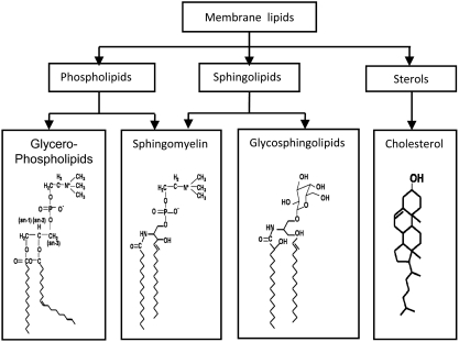
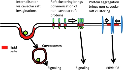
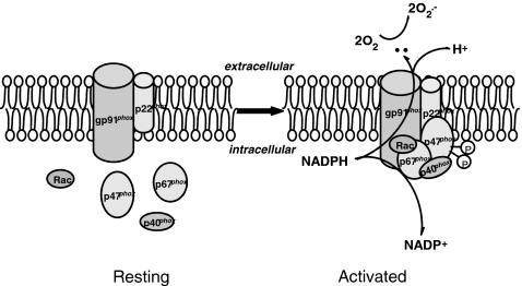
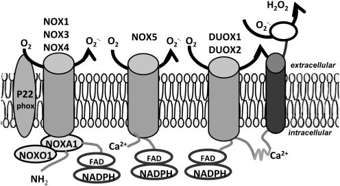
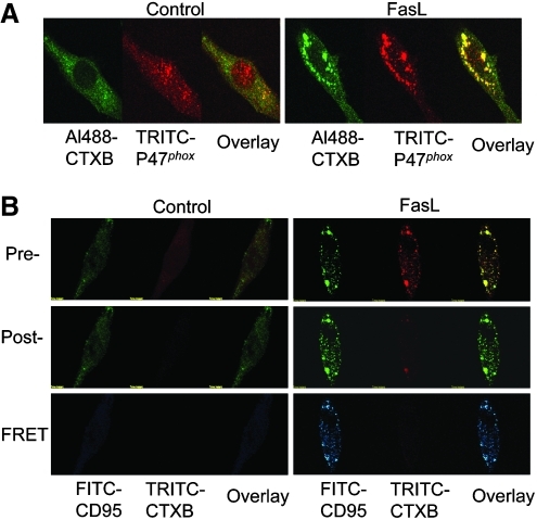
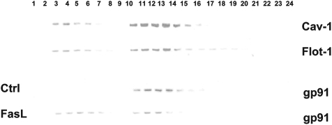
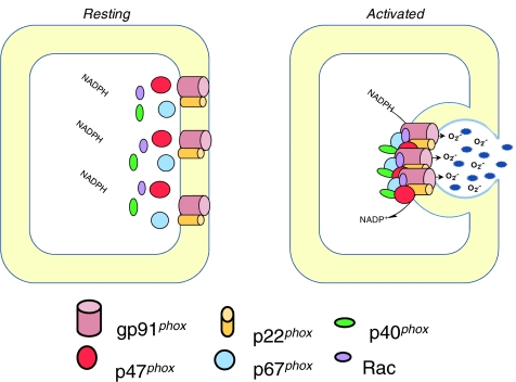
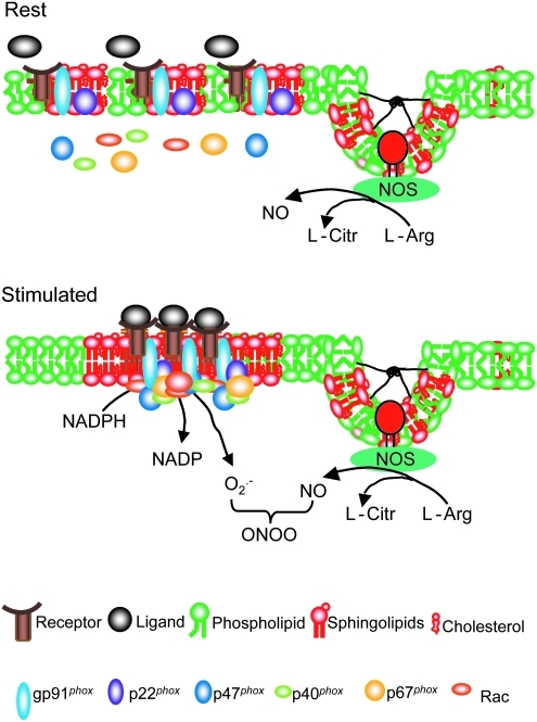
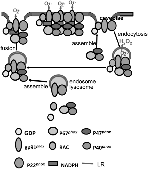
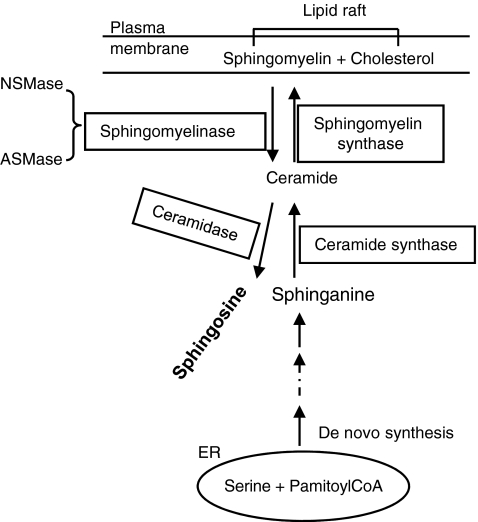
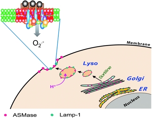
Lipid rafts, the sphingolipid and cholesterol-enriched membrane microdomains, are able to form different membrane macrodomains or platforms upon stimulations, including redox signaling platforms, which serve as a critical signaling mechanism to mediate or regulate cellular activities or functions. In particular, this raft platform formation provides an important driving force for the assembling of NADPH oxidase subunits and the recruitment of other related receptors, effectors, and regulatory components, resulting, in turn, in the activation of NADPH oxidase and downstream redox regulation of cell functions. This comprehensive review attempts to summarize all basic and advanced information about the formation, regulation, and functions of lipid raft redox signaling platforms as well as their physiological and pathophysiological relevance. Several molecular mechanisms involving the formation of lipid raft redox signaling platforms and the related therapeutic strategies targeting them are discussed. It is hoped that all information and thoughts included in this review could provide more comprehensive insights into the understanding of lipid raft redox signaling, in particular, of their molecular mechanisms, spatial-temporal regulations, and physiological, pathophysiological relevances to human health and diseases.


Figures

FIG. 1.
FIG. 1.
Composition of membrane lipids and their chemical structures. Lipid rafts (LRs) may consist of dynamic assemblies of cholesterol and lipids with saturated fatty acid chains such as sphingolipids and glycosphingolipids in the exoplasmic leaflet of the membrane bilayer. In addition, phospholipids with saturated fatty acids and cholesterol in the inner leaflet. Here depicted are structures of two sphingolipids including sphingomyelin and glycosphingolipids (GSL), cholesterol, and phospholipid-phosphatidylcholine.
FIG. 2.
FIG. 2.
Demonstration of caveolar and noncaveolar lipid rafts and their function. Caveolar and noncaveolar LRs may mediate different signaling pathways in different cells or even in the same cell in response to different agonists or stimuli. (To see this illustration in color the reader is referred to the web version of this article at www.liebertonline.com/ars).
FIG. 3.
FIG. 3.
Assembling and activation of NADPH oxidase. Upon stimulation, p47phox is phosphorylated and translocated to the membrane. NADPH oxidase subunits are aggregated in the membrane to form a functional enzyme. The gp91phox with help of other subunits or factors uses NADPH as substrate to transfer two electrons to molecular oxygen on the opposite side of the membrane to produce O2•−.
FIG. 4.
FIG. 4.
Major Nox isoforms and their proposed model of activation. In comparison, different NOXs may work in the same way as phagocytic NOX, which need the assembly of all subunits and cofactors, or in different way as phagocytic NOX, which nonphagocytic NOXs may be functioning without assembling other subunits or cofactors.
FIG. 5.
FIG. 5.
Confocal microscopic colocalization and FRET detection. (A) Colocalization of CTXB and p47phox as indicated by yellow spots or dots in overlay image, suggesting LR platforms or clusters. (B) The FRET as indicated by FITC-CD95 image and overlay image in blue. CD95 (Fas) is a typical LR-clustered receptor that activates LR clustering and redox signaling in ECs. (To see this illustration in color the reader is referred to the web version of this article at www.liebertonline.com/ars).
FIG. 6.
FIG. 6.
Flotation of membrane MR fractions by nondetergent 4- layer gradient flotation. A typical gel document shows that among 24 fractions, 3–6 and 10–14 are light and heavy low-density fractions, respectively. They represent noncaveolar and caveolar LRs. Under control condition, gp91phox was seen in caveolar fractions, but not in noncaveolar fractions. When the cells were treated with Fas ligand, the fractions were shifted to noncaveolar fractions.
FIG. 7.
FIG. 7.
LR-associated NADPH oxidase in respiratory burst. During phagocytic uptake of pathogens, LRs cluster, assemble, and activate NADPH oxidase in neutrophils to produce O2•−, causing respiratory burst. (To see this illustration in color the reader is referred to the web version of this article at www.liebertonline.com/ars).
FIG. 8.
FIG. 8.
LR redox signaling platforms associated with NADPH oxidase in transmembrane signaling. Under resting condition without ligand binding, individual LR with or without NOX are present in the membrane of ECs. When receptor ligand binding occurs, LRs are clustered to form LR platforms, with aggregation and assembling of NADPH oxidase subunits and other proteins such as Rac GTPase. Then, NADPH oxidase is activated to produce O2•−, which reacts with NO to produce ONOO, resulting in endothelial dysfunction. (To see this illustration in color the reader is referred to the web version of this article at www.liebertonline.com/ars).
FIG. 9.
FIG. 9.
Three models of LR redox signaling platforms. (i) LR redox signaling platforms is formed by clustering of LRs in plasma membrane upon stimulations of receptors to produce O2•− extracellularly or intracellularly. (ii) Upon stimulations, endocytosis occurs via caveolae to form intracellular LR-containing redoxosomes, producing O2•− and other reactive oxygen species to conduct redox signaling. (iii) During clustering of LRs, NOX and related subunits or cofactors are aggregated and then traffic to the plasma membrane to produce O2•− and conduct redox signaling.
FIG. 10.
FIG. 10.
Ceramide-metabolizing pathways. The de novo synthesis of ceramide begins with the conversion of dihydro-sphingosine into dihydro-ceramide by ceramide synthase and then further converted into ceramide. ASMase and NSMase catalyze the hydrolysis of phosphodiester bond in SM and produces ceramide and choline phosphate. Ceramide is converted back into SM by SM synthase and degraded by ceramidase.
FIG. 11.
FIG. 11.
Lysosome biogenesis and fusion to cell membrane to form LR platforms. ASMase is synthesized from the ER and transported through Golgi apparatus to lysosomes. These lysosomes can be mobilized to traffic and fuse into cell membrane, where ASMase is activated and ceramide produced, resulting in LRs clustering and formation of ceramide-enriched platforms. The insert presents an LR redox signaling platform or redox signalosome on cell membrane after lysosome fusion and activation of NADPH oxidase. (To see this illustration in color the reader is referred to the web version of this article at www.liebertonline.com/ars).

References

    1. Abdel Shakor AB. Kwiatkowska K. Sobota A. Cell surface ceramide generation precedes and controls FcgammaRII clustering and phosphorylation in rafts. J Biol Chem. 2004;279:36778–36787. - PubMed
    1. Abrahamsen H. Baillie G. Ngai J. Vang T. Nika K. Ruppelt A. Mustelin T. Zaccolo M. Houslay M. Tasken K. TCR- and CD28-mediated recruitment of phosphodiesterase 4 to lipid rafts potentiates TCR signaling. J Immunol. 2004;173:4847–4858. - PubMed
    1. Adam D. Heinrich M. Kabelitz D. Schutze S. Ceramide: does it matter for T cells? Trends Immunol. 2002;23:1–4. - PubMed
    1. Adam RM. Yang W. Di Vizio D. Mukhopadhyay NK. Steen H. Rapid preparation of nuclei-depleted detergent-resistant membrane fractions suitable for proteomics analysis. BMC Cell Biol. 2008;9:30. - PMC - PubMed
    1. Ago T. Kitazono T. Kuroda J. Kumai Y. Kamouchi M. Ooboshi H. Wakisaka M. Kawahara T. Rokutan K. Ibayashi S. Iida M. NAD(P)H oxidases in rat basilar arterial endothelial cells. Stroke. 2005;36:1040–1046. - PubMed

Publication types

LinkOut - more resources