. 2011 Feb 4;13(1):R14.
doi: 10.1186/ar3238.

Dual role of interleukin-17 in pannus growth and osteoclastogenesis in rheumatoid arthritis

Affiliations

Dual role of interleukin-17 in pannus growth and osteoclastogenesis in rheumatoid arthritis

Hiroshi Ito et al. Arthritis Res Ther. .

Abstract

Introduction: In a murine model, interleukin (IL)-17 plays a critical role in the pathogenesis of arthritis. There are controversies, however, regarding whether IL-17 is a proinflammatory mediator in rheumatoid arthritis (RA). We previously established an ex vivo cellular model using synovial tissue (ST)-derived inflammatory cells, which reproduced pannus-like tissue growth and osteoclastic activity in vitro. Using this model, we investigated the effects of IL-17 on pannus growth and osteoclastogenesis in RA.

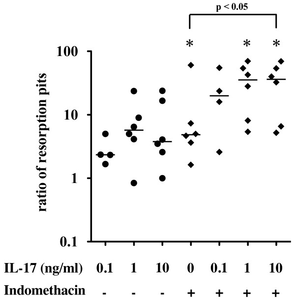
Methods: Inflammatory cells that infiltrated synovial tissue from patients with RA were collected without enzyme digestion and designated as ST-derived inflammatory cells. ST-derived inflammatory cells were cultured in the presence or absence of IL-17 or indomethacin, and the morphologic changes were observed for 4 weeks. Cytokines produced in the culture supernatants were measured by using enzyme-linked immunosorbent assay kits. Osteoclastic activity was assessed by the development of resorption pits in calcium phosphate-coated slides.

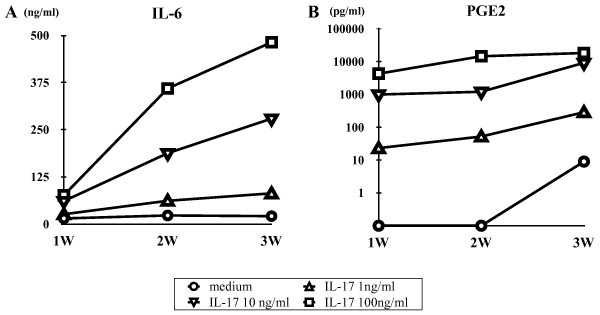
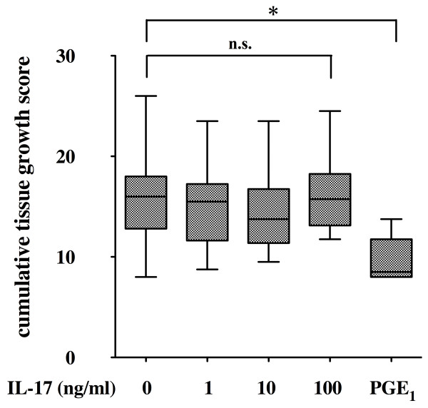
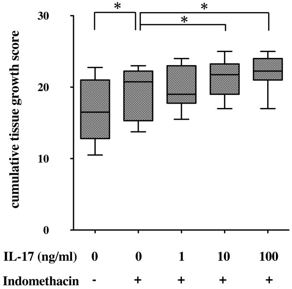
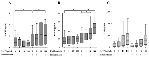
Results: Exogenous addition of IL-17 dramatically enhanced the spontaneous production of IL-6 and prostaglandin E₂ (PGE₂) by the ST-derived inflammatory cells, while it had no effect on the production of tumor necrosis factor (TNF)-α and macrophage colony-stimulating factor (M-CSF). Furthermore, IL-17 did not affect the spontaneous development of pannus-like tissue growth and osteoclastic activity by the ST-derived inflammatory cells. On the other hand, IL-17 enhanced pannus-like tissue growth, the production of TNF-α and M-CSF and the development of osteoclastic activity in the presence of indomethacin, an inhibitor of endogenous prostanoid production, while exogenous addition of PGE₁ suppressed their activities.

Conclusions: The present study suggests that IL-17 induces negative feedback regulation through the induction of PGE₂, while it stimulates proinflammatory pathways such as inflammatory cytokine production, pannus growth and osteoclastogenesis in RA.


Figures

Figure 1
Figure 1
Effect of interleukin (IL)-17 on the production of (A) IL-6 (A) and (B) prostaglandin E2 (PGE2) by the synovial tissue (ST)-derived inflammatory cells. Cells were incubated in the absence or presence of increasing concentrations of IL-17 (0 to 100 ng/ml) for 3 weeks. IL-6 (n = 7) and PGE2 (n = 3) in the culture supernatants were measured by enzyme-linked immunosorbent assay as described in Materials and methods.
Figure 2
Figure 2
Effect of interleukin (IL)-17 and prostaglandin E1 (PGE1) on pannus-like tissue growth in vitro. Synovial tissue (ST)-derived inflammatory cells were incubated in the absence or presence of increasing concentrations of IL-17 (0 to 100 ng/ml) (n = 17) or PGE1 (100 nM) (n = 9). Morphologic changes were observed under an inverted phase contrast microscope twice weekly for 4 weeks and were scored semiquantitatively on a scale of 0 to 4 according to the degree of tissue development as described in Materials and methods. Box and whisker plots indicate 25th/75th percentile and minimum/maximum, respectively. Bar indicates the median value. *P < 0.001 (by Mann-Whitney U test).
Figure 3
Figure 3
Effect of interleukin (IL)-17 on pannus-like tissue growth in the presence of indomethacin. Synovial tissue (ST)-derived inflammatory cells (n = 7) were incubated with an incremental dose of IL-17 in the absence or presence of indomethacin (100 to 1000 nM). Morphologic changes were observed under an inverted phase contrast microscope twice weekly for 4 weeks and were scored semiquantitatively on a scale of 0 to 4 according to the degree of tissue development as described in Materials and methods. Box and whisker plots indicate 25th/75th percentile and minimum/maximum, respectively. Bar indicates the median value. *P < 0.05 (by Wilcoxon signed-rank test).
Figure 4
Figure 4
Effect of interleukin (IL)-17 on the production of macrophage colony-stimulating factor (M-CSF), tumor necrosis factor α (TNF-α) and IL-6. Synovial tissue (ST)-derived inflammatory cells were incubated with incremental doses of IL-17 in the absence or presence of indomethacin (100 to 1,000 nM) for 1 week. Enzyme-linked immunosorbent assay kits were used to measure the concentration of (A) TNF-α, (B) M-CSF and IL-6 (C) in the culture supernatants derived from seven donors. There were no significant differences in the production of IL-6 between the presence and absence of indomethacin. Box and whisker plots indicate 25th/75th percentile and minimum/maximum, respectively. Bar indicates the median value. *P < 0.05 (by Wilcoxon signed-rank test).
Figure 5
Figure 5
Effect of interleukin (IL)-17 on the osteoclastogenesis. Osteoclastic activity was assessed by the development of resorption pits in calcium phosphate-coated slides as described in Materials and methods. Synovial tissue (ST)-derived inflammatory cells (n = 6) were cultured with incremental doses of IL-17 in the absence or presence of indomethacin on calcium phosphate-coated slides for 2 weeks and examined for the development of resorption pits. The ratio to the number of resorption pits in medium alone was plotted. Bar indicates the median value. *P < 0.05 vs. medium alone (by Wilcoxon signed-rank test).

References

    1. Chu CQ, Field M, Feldmann M, Maini RN. Localization of tumor necrosis factor α in synovial tissues and at the cartilage-pannus junction in patients with rheumatoid arthritis. Arthritis Rheum. 1991;34:1125–1132. doi: 10.1002/art.1780340908. - DOI - PubMed
    1. Buchan G, Barrett K, Turner M, Chantry D, Maini RN, Feldmann M. Interleukin-1 and tumour necrosis factor mRNA expression in rheumatoid arthritis: prolonged production of IL-1α. Clin Exp Immunol. 1988;73:449–455. - PMC - PubMed
    1. Hirano T, Matsuda T, Turner M, Miyasaka N, Buchan G, Tang B, Sato K, Shimizu M, Maini R, Feldmann M, Kishimoto T. Excessive production of interleukin 6/B cell stimulatory factor-2 in rheumatoid arthritis. Eur J Immunol. 1988;18:1797–1801. doi: 10.1002/eji.1830181122. - DOI - PubMed
    1. Bromley M, Woolley DE. Histopathology of the rheumatoid lesion. Identification of cell types at sites of cartilage erosion. Arthritis Rheum. 1984;27:857–863. doi: 10.1002/art.1780270804. - DOI - PubMed
    1. Maini R, St Clair EW, Breedveld F, Furst D, Kalden J, Weisman M, Smolen J, Emery P, Harriman G, Feldmann M, Lipsky P. Infliximab (chimeric anti-tumour necrosis factor α monoclonal antibody) versus placebo in rheumatoid arthritis patients receiving concomitant methotrexate: a randomised phase III trial. ATTRACT Study Group. Lancet. 1999;354:1932–1939. doi: 10.1016/S0140-6736(99)05246-0. - DOI - PubMed

Publication types

MeSH terms