. 2011 Feb 4;6(1):14.
doi: 10.1186/1750-1326-6-14.

Inhibition of RhoA GTPase and the subsequent activation of PTP1B protects cultured hippocampal neurons against amyloid β toxicity

Affiliations

Inhibition of RhoA GTPase and the subsequent activation of PTP1B protects cultured hippocampal neurons against amyloid β toxicity

Pedro J Chacon et al. Mol Neurodegener. .

Abstract

Background: Amyloid beta (Aβ) is the main agent responsible for the advent and progression of Alzheimer's disease. This peptide can at least partially antagonize nerve growth factor (NGF) signalling in neurons, which may be responsible for some of the effects produced by Aβ. Accordingly, better understanding the NGF signalling pathway may provide clues as to how to protect neurons from the toxic effects of Aβ.

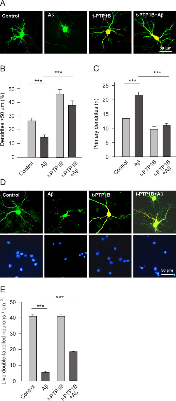
Results: We show here that Aβ activates the RhoA GTPase by binding to p75NTR, thereby preventing the NGF-induced activation of protein tyrosine phosphatase 1B (PTP1B) that is required for neuron survival. We also show that the inactivation of RhoA GTPase and the activation of PTP1B protect cultured hippocampal neurons against the noxious effects of Aβ. Indeed, either pharmacological inhibition of RhoA with C3 ADP ribosyl transferase or the transfection of cultured neurons with a dominant negative form of RhoA protects cultured hippocampal neurons from the effects of Aβ. In addition, over-expression of PTP1B also prevents the deleterious effects of Aβ on cultured hippocampal neurons.

Conclusion: Our findings indicate that potentiating the activity of NGF at the level of RhoA inactivation and PTP1B activation may represent a new means to combat the noxious effects of Aβ in Alzheimer's disease.


Figures

Figure 1
Figure 1
TAT-pep5 specifically uncouples p75NTR from the RhoA GTPase, and it counteracts the effects of Aβ on dendrite patterning, gene expression and the survival of cultured hippocampal neurons. (A, B and C) E17 hippocampal neurons were plated at a density of 40,000 cells/cm2 and cultured for 7 DIV. Neurons were transfected with pEGFP and then exposed for a further 16 h to TAT-Pep5 (1.0 μM), Aβ (5 μM), or both. The cells were fixed and labelled with the anti-EGFP antibody, and then processed for immunofluorescence. (A) representative micrographs of cultured neurons under the different conditions. The relative dendrite length (B) and primary dendrite numbers (C) was quantified as indicated in the methods section. Note that TAT-pep5 reversed the effects of Aβ on dendrite length and number. (D) Neurons cultured for 7 DIV (40,000 cells/cm2) were first incubated with TAT-Pep5 (1.0 μM) for 18 h, after which they were stimulated with Aβ (5 μM) for 4 h, lysed and then processed for real time PCR to quantify Hes1 expression. Note that TAT-pep5 prevented the Aβ-induced decrease in Hes1 mRNA. (E) 7 DIV cultures (30,000 cells/cm2) were stimulated with TAT-pep5 (1.0 μM) and/or Aβ (5 μM) for 90 h. The neurons were then stained with DAPI and those with intact nuclei were counted. Note that TAT-pep5 rescued around half of the neurons from the deleterious effects of Aβ. (F) Representative micrographs of DAPI stained nuclei in cultured hippocampal neurons treated with Aβ, or with Aβ and TAT-pep5, the latter conferring resistance against Aβ.
Figure 2
Figure 2
Aβ activates RhoA thereby influencing neuron morphology. (A) Western blots showing the activated and total RhoA GTPase in extracts from cultured PC12 nnr5 cells stimulated with NGF (100 ng/ml) for 5 h at the times indicated, or with Aβ (5 μM). Note that in contrast to NGF, Aβ increased the levels of RhoA GTP. The quantification of RhoA GTP in the lower panel is an average from four independent experiments. (B) Representative micrographs of hippocampal neurons cultured for 7 DIV (40,000 cells/cm2), treated with Aβ (5 μM) and/or co-transfected with EGFP and a myc tagged RhoA N19 (a dn form of RhoA) for 16 h. (C, D) Quantification of relative dendrite length (C) and primary dendrite number (D) in the four conditions indicated. Note that the attenuation of RhoA GTPase activity counteracted the effects of Aβ on dendrite length and number. Also note in (C) that the attenuation of RhoA activity increased the length of dendrites per se. (E, F) Quantification of relative dendrite length (E) and primary dendrite number (F) in cultured neurons after addition of CNFy (200 ng/ml: a specific activator of RhoA) for 16 h. (G) 7 DIV neurons in culture were first incubated with C3 ADP rybosyl transferase (1.0 μM) for 18 h, they were stimulated with Aβ (5 μM) for 4 h, lysed and then processed for real time PCR to quantify Hes1 expression. Note that the inhibition of RhoA by C3 prevented the Aβ-induced decrease in Hes1 mRNA.
Figure 3
Figure 3
The role of RhoA in Aβ induced neuron death. (A, B) Hippocampal neurons (30,000 cells/cm2) were cultured for 7 days and then treated with Aβ (5 μM). Two days later the neurons were transfected with the dn RhoA N19 and on the following day, the cells were stained and the number of live cells were determined as described in the Methods. (A) Representative micrographs of double-labelled cultured hippocampal neurons under the four conditions described. Green represents EGFP immunostaining, red is the transfected myc-tagged RhoA N19 and the DAPI stained nuclei are blue. (B) Quantification of live cells. Note that transfection with the dominant negative form of RhoA rescued a significant number of neurons from Aβ-induced death. (C) The effects of Anti-amyloid were more dramatic when C3 ADP ribosyl transferase (1 μM), a RhoA inhibitor, was applied to the cultures. Cultured hippocampal neurons (7 DIV) were treated simultaneously with C3 ADP ribosyl transferase and Aβ, and the number of live cells was determined four days later in culture.
Figure 4
Figure 4
Amyloid β interferes with the capacity of NGF to activate protein tyrosine phosphatase 1B in hippocampal neurons. Cultured 7 DIV neurons (about 250,000 cells per experimental point) were treated as indicated and PTP1B activity was assessed. (A) Whereas NGF (100 ng/ml) increased PTP1B activity several fold, Aβ (5 μM) did not alter the activity of the phosphatase, although Aβ did prevent the increase of PTP1B activity induced by NGF (B). Another activator of RhoA activity, CNFy (200 ng/ml), also prevented the activation of PTP1B caused by NGF. Both Aβ and CNFy were applied to cultures 18 h before stimulating with NGF for 4 h. (C) The inhibition of RhoA activity after incubating the cells with either C3 (1 μM) or TAT-pep5 (1 μM) for 18 h increased the activity of PTP1B. By contrast to NGF, such increases were not counteracted by prior application of Aβ.
Figure 5
Figure 5
Overexpression of PTP1B counteracts the effects of Aβ on dendrite patterning (A, B, C) and neuron death (D, E). Cultured hippocampal neurons (40,000 cells/cm2, 7 DIV) were co-transfected with the EGFP and PTP1B plasmids, treated with Aβ (5 μM) and incubated for a further 16 h to analyse dendrite patterning (A, B, C). (A) Representative micrographs of cultured hippocampal neurons at 7 DIV treated with Aβ and/or transfected with PTP1B. EGFP immunostaining is in green and the transfected HA-tagged PTP1B in red. (B, C) Quantification of the relative dendrite length (B) and primary dendrite number (C) in the four conditions indicated. Note that overexpression of PTP1B increased dendrite length and prevented the morphological effects of Aβ. (D, E) Hippocampal neurons (30,000 cells/cm2) were cultured for 7 days and then treated with Aβ (5 μM). Two days later, the neurons were transfected with the PTP1B expressing plasmid, and on the following day the cells were stained and the live cells determined as described in the Methods. (D) Representative micrographs of double-labelled cultured hippocampal neurons under the four conditions described. EGFP immunostaining is in green, the transfected HA-tagged PTP1B in red and the DAPI stained nuclei are blue. (E) Quantification of live cells. Note that transfection with the PTP1B expressing plasmid rescued a significant number of neurons from Aβ-induced neuron death.
Figure 6
Figure 6
Diagram showing the signals transduced by NGF leading to Hes1 expression and neuron survival, and their impairment by Aβ. Various steps have been defined here and are shown in red (labelled as 2). The steps in black ((3), (4), (5) and (6)) come from the literature and the steps labelled in blue (1 and 7) are from our previous studies [19-21,29].

Similar articles

Cited by

References

    1. Wilcock DM, Colton CA. Anti-amyloid-beta immunotherapy in Alzheimer's disease: relevance of transgenic mouse studies to clinical trials. J Alzheimers Dis. 2008;15:555–569. - PMC - PubMed
    1. Nerelius C, Gustafsson M, Nordling K, Larsson A, Johansson J. Anti-amyloid activity of the C-terminal domain of proSP-C against amyloid beta-peptide and medin. Biochemistry. 2009;48:3778–3786. doi: 10.1021/bi900135c. - DOI - PubMed
    1. Stains CI, Mondal K, Ghosh I. Molecules that target beta-amyloid. ChemMedChem. 2007;2:1674–1692. doi: 10.1002/cmdc.200700140. - DOI - PubMed
    1. Foster JK, Verdile G, Bates KA, Martins RN. Immunization in Alzheimer's disease: naive hope or realistic clinical potential? Mol Psychiatry. 2009;14:239–251. doi: 10.1038/mp.2008.115. - DOI - PubMed
    1. St George-Hyslop PH, Morris JC. Will anti-amyloid therapies work for Alzheimer's disease? Lancet. 2008;372:180–182. doi: 10.1016/S0140-6736(08)61047-8. - DOI - PubMed