. 2011 Feb 4:9:22.
doi: 10.1186/1477-7827-9-22.

Modulation of small leucine-rich proteoglycans (SLRPs) expression in the mouse uterus by estradiol and progesterone

Affiliations

Modulation of small leucine-rich proteoglycans (SLRPs) expression in the mouse uterus by estradiol and progesterone

Renato M Salgado et al. Reprod Biol Endocrinol. .

Abstract

Background: We have previously demonstrated that four members of the family of small leucine-rich-proteoglycans (SLRPs) of the extracellular matrix (ECM), named decorin, biglycan, lumican and fibromodulin, are deeply remodeled in mouse uterine tissues along the estrous cycle and early pregnancy. It is known that the combined action of estrogen (E2) and progesterone (P4) orchestrates the estrous cycle and prepares the endometrium for pregnancy, modulating synthesis, deposition and degradation of various molecules. Indeed, we showed that versican, another proteoglycan of the ECM, is under hormonal control in the uterine tissues.

Methods: E2 and/or medroxiprogesterone acetate (MPA) were used to demonstrate, by real time PCR and immunoperoxidase staining, respectively, their effects on mRNA expression and protein deposition of these SLRPs, in the uterine tissues.

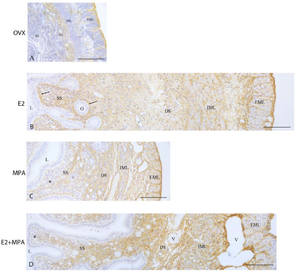
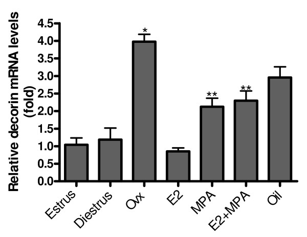
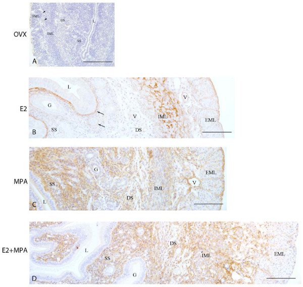
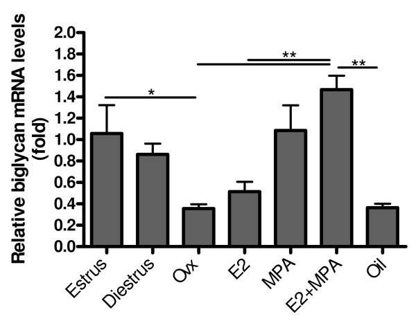
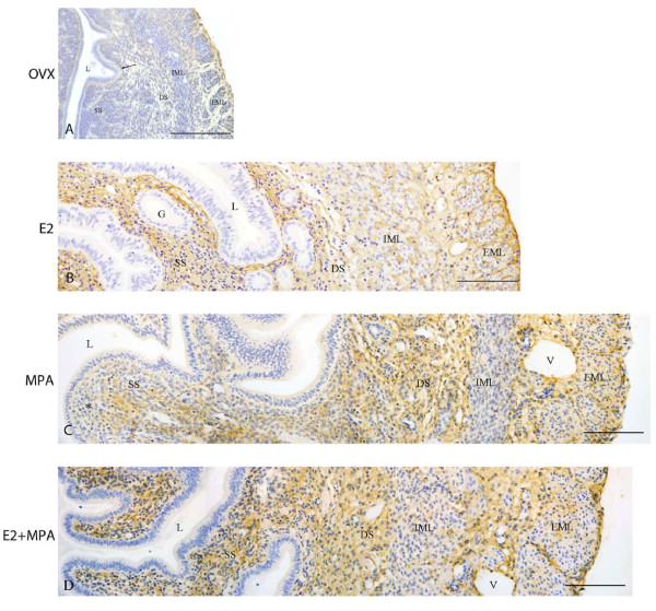
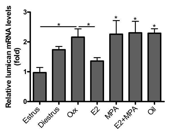
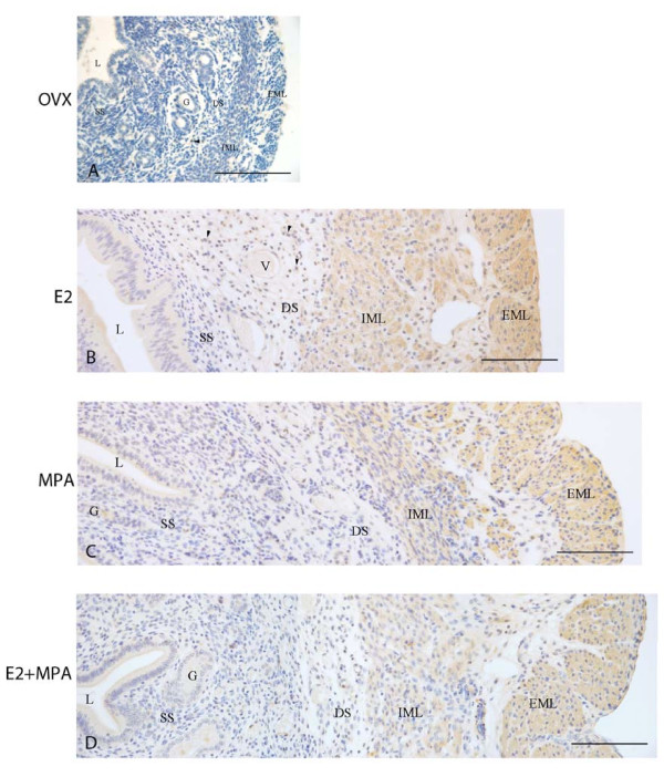
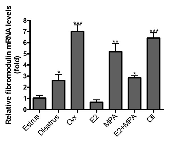
Results: Decorin and lumican were constitutively expressed and deposited in the ECM in the absence of the ovarian hormones, whereas deposition of biglycan and fibromodulin were abolished from the uterine ECM in the non-treated group. Interestingly, ovariectomy promoted an increase in decorin, lumican and fibromodulin mRNA levels, while biglycan mRNA conspicuously decreased. Hormone replacement with E2 and/or MPA differentially modulates their expression and deposition.

Conclusions: The patterns of expression of these SLRPs in the uterine tissues were found to be hormone-dependent and uterine compartment-related. These results reinforce the existence of subpopulations of endometrial fibroblasts, localized into distinct functional uterine compartments, resembling the organization into basal and functional layers of the human endometrium.


Figures

Figure 1
Figure 1
Immunoperoxidase for decorin. (A) Ovx group: decorin is present in the deep stroma, the external muscle layer and the connective tissue between layers; (B) E2-group: the immunoreaction is seen in the whole endometrial stroma, especially around glands and under the luminal epithelium (arrows), as well as in both layers of the myometrium; (C) MPA-group: the reaction is observed in the deep stroma and only traces are seen in the superficial stroma (asterisk). In the myometrium, the reaction is present in both layers and the connective tissue; (D) E2+MPA-group: the deposition of decorin is observed in the whole endometrial stroma and in both layers of the myometrium, as well ass in the connective tissue between them. L = uterine lumen; G = endometrial gland; SS = superficial stroma; DS = deep stroma; V = blood vessel; IML = internal muscle layer; EML = external muscle layer. Scale bar: 50 μm.
Figure 2
Figure 2
Decorin mRNA expression. Ovariectomy (ovx) significantly increases decorin mRNA expression (~4 fold) when compared with estrus. Hormone replacement promotes a significant reduction in decorin expression in the E2-treated group (~4 fold) and MPA and E2+MPA-treated groups (~2 fold). Values represent the mean ± SEM. *p < 0.05; **p < 0.01; ***p < 0.001.
Figure 3
Figure 3
Immunoperoxidase for biglycan. (A) Ovx group: the immunoreaction is absent in all uterine tissues, being detected only in the cytoplasm of some leukocytes (arrowhead); (B) E2-group: the reaction is observed in the region of basement membrane of the luminal and glandular epithelia (arrows) and in the myometrium; (C) MPA-group: strong diffuse reaction in the whole endometrial stroma. Moderate immunoreaction is present in both layers of the myometrium and the connective tissue between them; (D) E2+MPA-group: the immunoreaction is distributed as a network of thin filaments in the whole stroma. In the myometrium, the deposition of biglycan is observed in both layers and connective tissue, being weak in the EML. L = uterine lumen; G = endometrial gland; SS = superficial stroma; DS = deep stroma; V = blood vessel; IML = internal muscle layer; EML = external muscle layer. Scale bar: 50 μm.
Figure 4
Figure 4
Biglycan mRNA expression. After ovariectomy there is a drastic reduction in biglycan expression (~3 fold), when compared with estrus. A significant increase is observed after hormone treatment with MPA (~3 fold) and E2+MPA (~5 fold). Values represent the mean ± SEM. *p < 0.05; **p < 0.01; ***p < 0.001.
Figure 5
Figure 5
Immunoperoxidase for lumican. (A) Ovx group: A weak immunoreaction is observed in the endometrial stroma, especially under the luminal epithelium (arrow) and in the deep stroma, as well as in the EML and connective tissue between muscle layers of the myometrium; (B) E2-group: the reaction is present in the whole endometrial stroma and in both layers of the myometrium; (C) MPA-group: the reaction is present in the whole stroma, however it is weak in the subepithelial stroma (asterisk). In the myometrium, deposition of lumican is observed in the EML and connective tissue between layers; (D) E2+MPA-group: the immunoreaction is strong in the whole endometrial stroma. In the myometrium, a weak staining is observed in both IML and EML, being strong in the connective tissue between them. L = uterine lumen; G = endometrial gland; SS = superficial stroma; DS = deep stroma; V = blood vessel; IML = internal muscle layer; EML = external muscle layer. Scale bar: 50 μm.
Figure 6
Figure 6
Lumican mRNA expression. After ovariectomy there is a ~2 fold increase in lumican mRNA levels, when compared with estrus. Hormone treatments do not significantly alter lumican expression in the uterus. Values represent the mean ± SEM. *p < 0.05; **p < 0.01; ***p < 0.001.
Figure 7
Figure 7
Immunoperoxidase for fibromodulin. (A) Ovx group: the immunodeposition is absent from all uterine tissues, but may be observed in the cytoplasm of some leukocytes (arrowhead); (B) E2-group: the immunoreaction is present in the apical cytoplasm of epithelial cells and in leukocytes observed in the stroma (arrowheads). A strong immunoreaction is seen in both layers of the myometrium and in the connective tissue between them; (C) MPA-group: the reaction is present in the luminal epithelium and in the secretion of endometrial glands. The immunoreaction is seen in both layers of the myometrium, especially the EML; (D) E2+MPA-group: the staining pattern is similar to that observed in the MPA group. L = uterine lumen; G = endometrial gland; SS = superficial stroma; DS = deep stroma; V = blood vessel; IML = internal muscle layer; EML = external muscle layer. Scale bar: 50 μm.
Figure 8
Figure 8
Fibromodulin mRNA expression. The expression is significantly higher in diestrus than in estrus (~2 fold). Moreover, ovariectomy promotes a conspicuous increase in fibromodulin mRNA levels, when compared to estrus (~7 fold). Hormone treatments differentially induce decrease in fibromodulin expression (E2 - ~7 fold; MPA - ~2 fold; E2+MPA - ~2.5 fold). Values represent the mean ± SEM. *p < 0.05; **p < 0.01; ***p < 0.001.

Similar articles

Cited by

References

    1. Allen E. The oestrous cycle in the mouse. Am J Anat. 1927;30:297–371. doi: 10.1002/aja.1000300303. - DOI
    1. Martin L, Finn CA. Hormone secretion during early pregnancy in the mouse. J Endocrinol. 1969;45:57–65. doi: 10.1677/joe.0.0450057. - DOI - PubMed
    1. Parandoosh Z, Crombie DL, Tetzke TA, Hayes JS, Heap RB, Wang M-W. Progesterone and oestrogen receptors in the decidualized mouse uterus and effects of different types of anti-progesterone treatment. J Reprod Fertil. 1995;105:215–220. doi: 10.1530/jrf.0.1050215. - DOI - PubMed
    1. Lydon JP, DeMayo FJ, Funk CR, Mani SK, Hughes AR, Montgomery CA Jr, Shyamala G, Conneely OM, O'Malley BW. Mice lacking progesterone receptor exhibit pleiotropic reproductive abnormalities. Genes Dev. 1995;9:2266–2278. doi: 10.1101/gad.9.18.2266. - DOI - PubMed
    1. Weihua Z, Saji S, Sirpa M, Cheng G, Jensen EV, Warner M, Gustafsson J. Estrogen receptor (ER) β, a modulator of ERα in the uterus. Procl Natl Acad Sci USA. 2000;97:5936–5941. doi: 10.1073/pnas.97.11.5936. - DOI - PMC - PubMed

Publication types

LinkOut - more resources