

Dermatofibrosarcoma protuberans with fibrosarcomatous transformation of the head and neck
- PMID: 21294902
- PMCID: PMC3038985
- DOI: 10.1186/1758-3284-3-5
Dermatofibrosarcoma protuberans with fibrosarcomatous transformation of the head and neck
Abstract
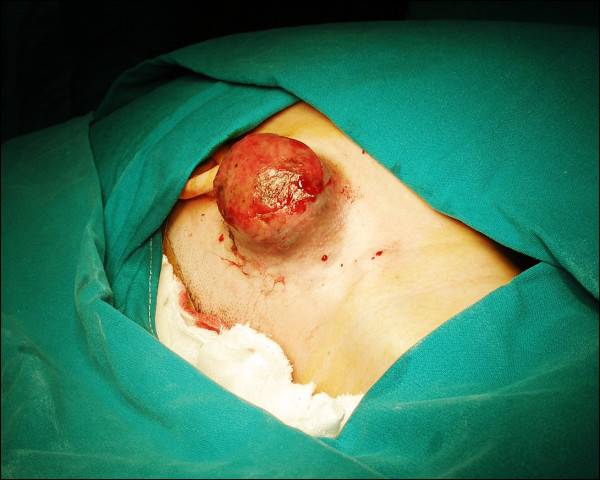
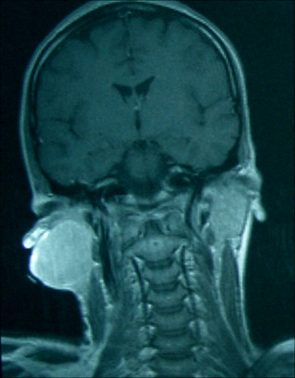
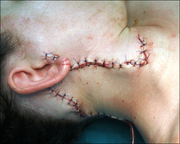
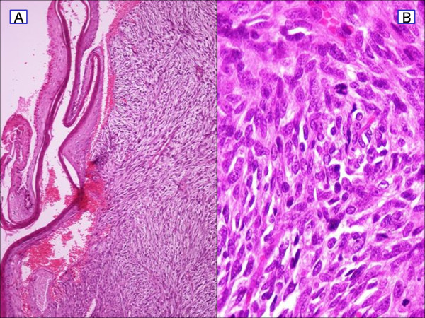
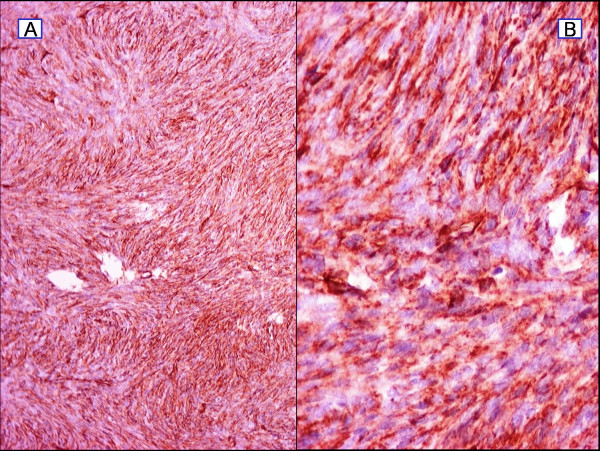
Dermatofibrosarcoma protuberans (DFSP) is a rare cutaneous neoplasm associated with a high cure rate. We present a case of aggressive DFSP with fibrosarcomatous areas in the head and neck. A 28-year-old Mediterranean female presented with a 45-day history of rapidly growing cutaneous lesion of the face. Surgical biopsy confirmed the diagnosis of DFSP. Subsequently, the patient underwent wide local surgical resection, followed by reconstruction. Histopathology report revealed fibrosarcomatous transformation and the patient underwent adjuvant radiotherapy. The patient continues to be disease free at the 35-month follow-up.Although DFSP behave as non-aggressive malignancy, surgery with complete removal of the affected area is the intervention of choice. Moreover, adjuvant treatment and follow-up of the patient is essential in order to prevent recurrence.
Figures

References
-
- Taylor RW. Sarcomatous tumors resembling in some respects keloids. Arch Dermatol. 1890;8:384–387.
-
- Darrier J, Ferrand M. Dermatofibromes progressifs et recidivants ou fibrosarcomes de la peau. Ann Dermatol Syphiligr (Paris) 1924;5:545–62.
-
- Hoffman E. Uber das knollentreibende fibrosarkom der haut. Dermatol Zischr. 1925;43:1–28. doi: 10.1159/000250699. - DOI
Publication types
MeSH terms
LinkOut - more resources
Full Text Sources
Medical
