

Enhancive effects of Lewis y antigen on CD44-mediated adhesion and spreading of human ovarian cancer cell line RMG-I
- PMID: 21294926
- PMCID: PMC3045975
- DOI: 10.1186/1756-9966-30-15
Enhancive effects of Lewis y antigen on CD44-mediated adhesion and spreading of human ovarian cancer cell line RMG-I
Abstract
Background: This study aimed to investigate the molecular structural relationship between cell adhesive molecule CD44 and Lewis y antigen, and determine the effects of Lewis y antigen on CD44-mediated adhesion and spreading of ovarian cancer cell line RMG-I and the Lewis y antigen-overexpressed cell line RMG-I-H.
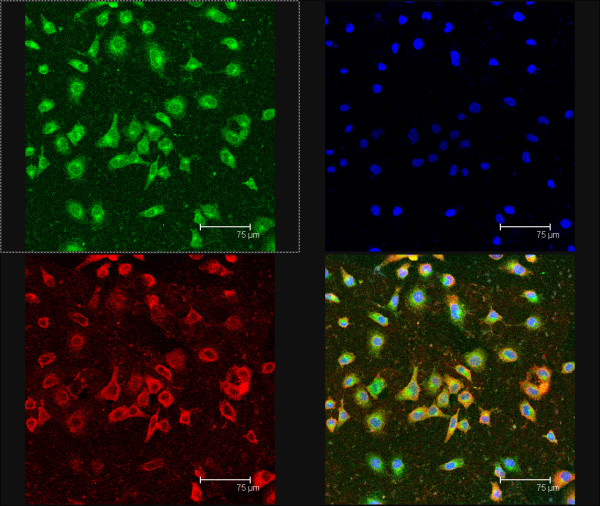
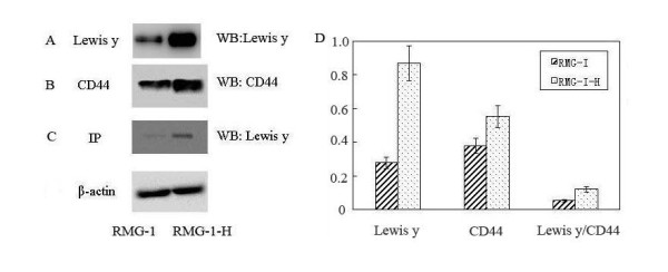
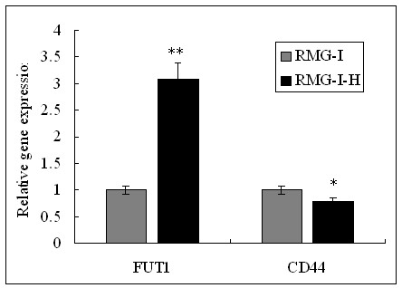
Methods: The expression of CD44 in RMG-I and RMG-I-H cells before and after treatment of Lewis y monoclonal antibody was detected by immunocytochemistry; the expression of Lewis y antigen and CD44 was detected by Western Blot. The structural relationship between Lewis y antigen and CD44 was determined by immunoprecipitation and confocal laser scanning microscopy. The adhesion and spreading of RMG-I and RMG-I-H cells on hyaluronic acid (HA) were observed. The expression of CD44 mRNA in RMG-I and RMG-I-H cells was detected by real-time RT-PCR.
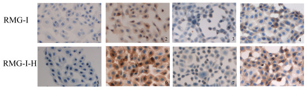
Results: Immunocytochemistry revealed that the expression of CD44 was significantly higher in RMG-I-H cells than in RMG-I cells (P < 0.01), and its expression in both cell lines was significantly decreased after treatment of Lewis y monoclonal antibody (both P < 0.01). Western Blot confirmed that the content of CD44 in RMG-I-H cells was 1.46 times of that in RMG-I cells. The co-location of Lewis y antigen and CD44 was confirmed by co-immunoprecipitation. The co-expression of CD44 and Lewis y antigen in RMG-I-H cells was 2.24 times of that in RMG-I cells. The adhesion and spreading of RMG-I-H cells on HA were significantly enhanced as compared to those of RMG-I cells (P < 0.01), and this enhancement was inhibited by Lewis y monoclonal antibody (P < 0.01). The mRNA level of CD44 in both cell lines was similar (P > 0.05).
Conclusion: Lewis y antigen strengthens CD44-mediated adhesion and spreading of ovarian cancer cells.
Figures
References
-
- Ugorski M, Laskowska A. Sialyl Lewis a: a tumor-associated carbohydrate antigen involved in adhesion and metastatic potential of cancer cells. Acta Biochim Pol. 2002;49:303–311. - PubMed
-
- Diao B, Lin B. Lewis y antigen and its applications to tumor diagnosis and treatment. J Modern Oncol. 2009;17:132–134.
-
- Rodríguez-Burford C, Barnes MN, Berry W, Partridge EE, Grizzle WE. Immunohistochemical expression of molecular markers in an avian model: a potential model for preclinical evaluation of agents for ovarian cancer chemoprevention. Gynecol Oncol. 2001;81:373–379. - PubMed
-
- Hao YY, Lin B, Zhao Y, Zhang YH, Li FF, Diao B, Ou YL, Zhang SL. α1, 2-Fucosyltransferase gene transfection influences on biological behavior of ovarian carcinoma-derived RMG-I cells. Fen Zi Xi Bao Sheng Wu Xue Bao. 2008;41:435–442. - PubMed
-
- Iwamori M, Tanaka K, Kubushiro K, Lin B, Kiguchi K, Ishiwata I, Tsukazaki K, Nozawa S. Alterations in the glycolipid composition and cellular properties of ovarian carcinoma-derived RMG-I cells on transfecton of the alpha 1,2-fucosyltransferase gene. Cancer Sci. 2005;96:26–30. doi: 10.1111/j.1349-7006.2005.00005.x. - DOI - PMC - PubMed
Publication types
MeSH terms
Substances
LinkOut - more resources
Full Text Sources
Medical
Miscellaneous
