. 2011 Apr 1;407(3):439-49.
doi: 10.1016/j.jmb.2011.01.057. Epub 2011 Feb 3.

Non-proteolytic functions of calpain-3 in sarcoplasmic reticulum in skeletal muscles

Affiliations

Non-proteolytic functions of calpain-3 in sarcoplasmic reticulum in skeletal muscles

Koichi Ojima et al. J Mol Biol. .

Abstract

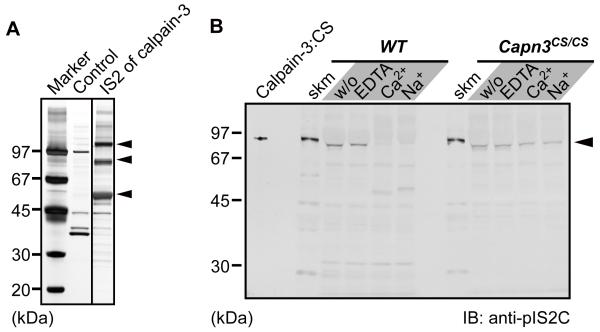
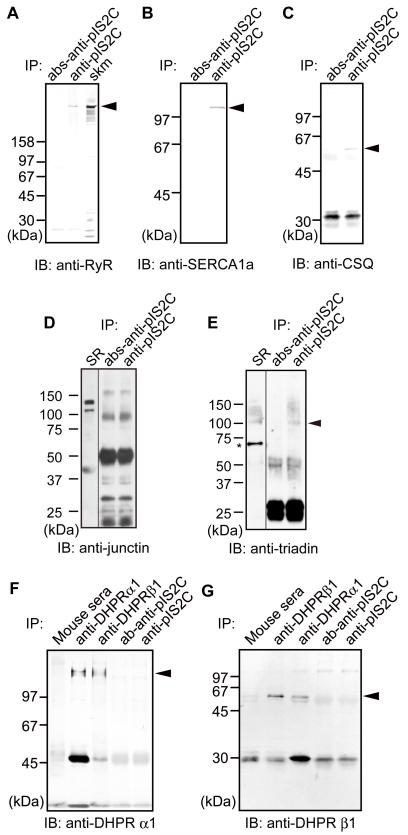
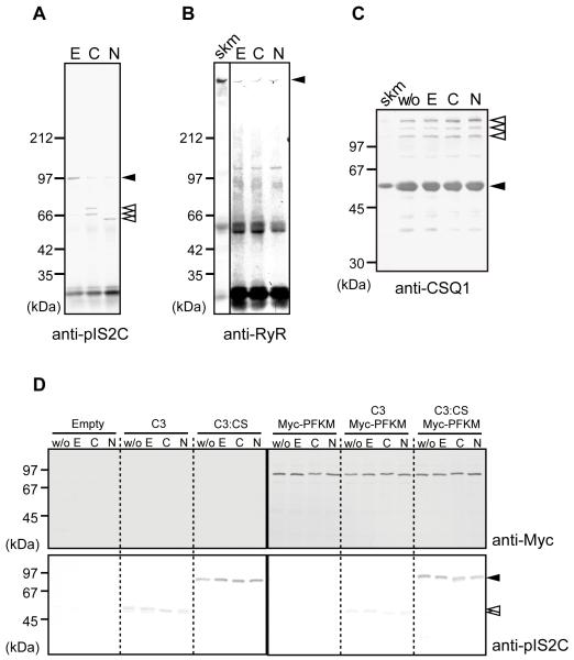
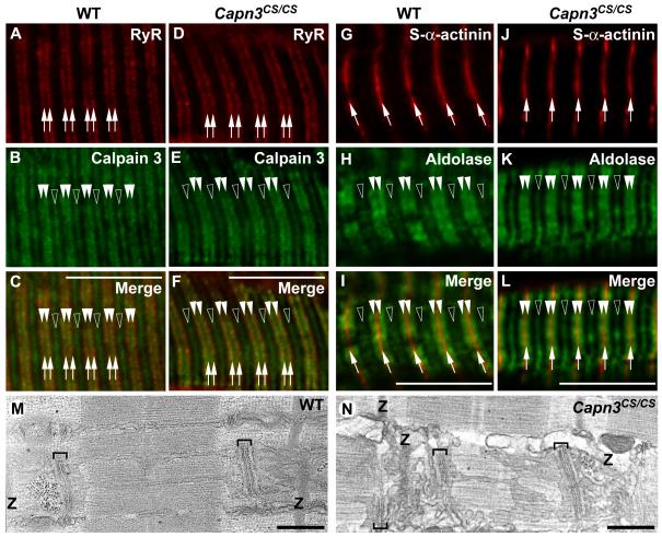
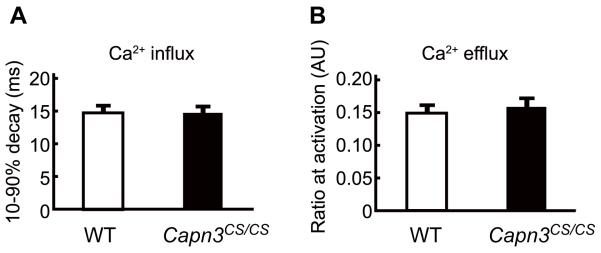
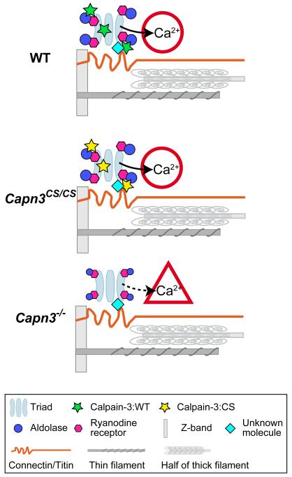
Mutations in CAPN3/Capn3, which codes for skeletal muscle-specific calpain-3/p94 protease, are responsible for limb-girdle muscular dystrophy type 2A. Using "knock-in" (referred to as Capn3(CS/CS)) mice, in which the endogenous calpain-3 is replaced with a mutant calpain-3:C129S, which is a proteolytically inactive but structurally intact calpain-3, we demonstrated in our previous studies that loss of calpain-3 protease activity causes muscular dystrophy [Ojima, K. et al. (2010) J. Clin. Invest. 120, 2672-2683]. However, compared to Capn3-null (Capn3(-/-)) mice, Capn3(CS/CS) mice showed less severe dystrophic symptoms. This suggests that calpain-3 also has a non-proteolytic function. This study aimed to elucidate the non-proteolytic functions of calpain-3 through comparison of Capn3(CS/CS) mice with Capn3(-/-) mice. We found that calpain-3 is a component of the sarcoplasmic reticulum (SR), and that calpain-3 interacts with, but does not proteolyze, typical SR components such as ryanodine receptor and calsequestrin. Furthermore, Capn3(CS/CS) mice showed that the nonenzymatic role of calpain-3 is required for proper Ca(2+) efflux from the SR to cytosol during muscle contraction. These results indicate that calpain-3 functions as a nonenzymatic element for the Ca(2+) efflux machinery in the SR, rather than as a protease. Thus, defects in the nonenzymatic function of calpain-3 must also be involved in the pathogenesis of limb-girdle muscular dystrophy type 2A.


Figures

Fig. 1
Fig. 1
Identification of calpain-3 IS2 region interacting molecules. (A) Identification of calpain-3-specific insertion 2 (IS2)-interacting proteins. Skeletal muscle lysates were pulled down with an IS2 peptide column, and subsequently the trapped proteins were electrophoresed and detected by silver staining. Three major bands at 110, 90, and 55 kDa (arrowheads) were identified as SERCA1a, phosphofructose kinase muscle type, and CSQ1, respectively, by MALDI-TOF/TOF analysis. (B) Immunoblot detection of calpain-3 in a microsomal fraction. Microsomal fractions isolated from WT and Capn3CS/CS skeletal muscles were incubated with either 10 mM EDTA, 5 mM CaCl2, or 150 mM NaCl, at 30°C for 0 min (w/o) or 30 min. calpain-3:CS indicates the lysate from COS7 cells exogenously expressing human calpain-3:C129S. Slightly faster migration of the calpain-3 bands in the microsomal fractions than in total muscle fractions (skm) is because a large amount of SERCA1a migrating just above calpain-3 on the gel pushed calpain-3 downwards.
Fig. 2
Fig. 2
Calpain-3 associates with protein complexes in the SR. IP of microsomal fractions with anti-pIS2C antibody analyzed by western blot using antibodies against RyR (A), SERCA1a (B), CSQ1(C), junctin (D), triadin (E), DHPRα1 (F) and DHPRβ1 (G). Peptide-absorbed anti-calpain-3 (pIS2C) antibody (abs-anti-pIS2C) and normal mouse sera were used as negative controls. Junctin is one of the isoforms of aspartyl/asparaginyl α-hydroxylase (ASPH). Apparent molecular weight of junctin is about 40,000, although its calculated molecular weight is 26,000. Closed arrowheads, indicated proteins; IB, immunoblot; skm, total muscle fractions as a positive control for RyR; SR, microsome fraction as a positive control for junctin and triadin; *non-specific band detected by anti-triadin antibody used.
Fig. 3
Fig. 3
Calpain-3 did not proteolyze molecules in the SR. (A-B) IPs of microsomal fractions from WT mice by anti-pIS2C antibody were incubated with 10 mM EDTA (E), 5 mM CaCl2 (C), or 150 mM NaCl (N), and detected by anti-IS2C (A) or anti-RyR (B). In the presence of Ca2+ or Na+, calpain-3 was autolyzed. Open arrowheads, autolytic fragments of calpain-3; closed arrowheads, full-length calpain-3 (A), and full-length RyR (B). No RyR fragments were observed (B). (C) The microsomal fraction from WT mice was treated with none (w/o), 10 mM EDTA (E), 5 mM CaCl2 (C), or 150 mM NaCl (N), and was subjected to western blot analysis using anti-CSQ1 antibody. No breakdown product for CSQ1 was observed. Closed arrowhead, CSQ1; open arrowheads, CSQ-like proteins; skm, total skeletal muscle lysate. (D) Lysates of COS7 cells transfected with empty pcDNA3.1 vector (Empty), or expression vectors for calpain-3 (C3), calpain-3:C129S (C3:CS), and/or Myc-PFKM, were incubated with 10 mM EDTA (E), 5 mM CaCl2 (C), or 150 mM NaCl (N). “w/o” indicates no incubation control. No proteolysis of Myc-PFKM was observed in any lane when detected with anti-Myc antibody. Calpain-3 was detected by anti-pIS2C antibody (closed and open arrowheads indicate full-length and autolytic fragments of calpain-3, respectively). Note that wild type calpain-3 autolyzed without incubation, but that further band shift was observed when incubated with CaCl2 or NaCl.
Fig. 4
Fig. 4
Localizations of RyR and aldolase A were not affected by a loss of calpain-3 protease activity. (A-L) Longitudinal cryostat sections of EDL from WT (A-C, G-I) and Capn3CS/CS (D-F, J-L) mice were reacted with antibodies against RyR (A, D), calpain-3 (B, E), s-α-actinin (G, J), and aldolase A (H, K). RyR (arrows in A, C, D, F) partially co-localized with calpain-3 in the N2A regions of both WT and Capn3CS/CS (closed arrowheads in B, C, E, F). In addition to the N2A regions, calpain-3 was detected in the M-lines as we previously reported (open arrowheads in B and E). Localizations of aldolase A in both WT and Capn3CS/CS showed the identical patterns, i.e. doublets along the Z-bands (closed arrowheads in G, I, J, L), and near the M-lines (open arrowheads in H, I, K, L). Bars, 10 μm. (M-N) Representative electron microscopic images of longitudinal sections of EDL from WT (M) and Capn3CS/CS (N) mice. The positions of the Z-bands in the sarcomeres are indicated as “Z”. Brackets indicate triads, in which a T-tubule is sandwiched between two junctional SR cisternae. The triad locates at the A-I junctions of the sarcomeres in both types of mice. The longitudinal SR runs in parallel with myofibrils between two triads. Bars, 0.5 μm.
Fig. 5
Fig. 5
Proteolytic function of Calpain-3 is not required for SR calcium kinetics. (A, B) Myofibers isolated from FDB muscle of WT and Capn3CS/CS mice were loaded with Fura2FF/AM, twitch activated and Ca2+ transients were measured. The time required for the decay of Fura2FF/AM fluorescence from 90 to 10% of its peak was used to determine the speed of Ca2+ influx from the cytosol to the SR (A). Ca2+ efflux from the SR to the cytosol was measured by the ratio of Ca2+ bound to Ca2+ unbound fluorescence upon myofiber stimulation (B). There was no significant difference in Ca2+ influx and speed of efflux between WT and Capn3CS/CS. The numbers of measured myofibers from WT and Capn3CS/CS were 24 and 22, respectively. Error bars, SE.
Fig. 6
Fig. 6
Model of calpain-3 structural functions in the SR. The triads and major myofibrillar components are shown in a half sarcomere. (A) In WT mice, calpain-3 molecules (with proteolytic activity suppressed by an unknown mechanism) are structurally associated with SR components, and normal Ca2+ efflux from the SR takes place. (B) In Capn3CS/CS mice, normal Ca2+ efflux from the SR also takes place because calpain-3:C129S is proteolytically inactive calpain-3 but structurally intact. (C) In Capn3−/− mice, however, the calpain-3 molecules is lost and as a result Ca2+ efflux from the SR is reduced.

References

    1. Goll DE, Thompson VF, Li H, Wei W, Cong J. The calpain system. Physiol. Rev. 2003;83:731–801. - PubMed
    1. Suzuki K, Hata S, Kawabata Y, Sorimachi H. Structure, activation, and biology of calpain. Diabetes. 2004;53:S12–18. - PubMed
    1. Sorimachi H, Imajoh-Ohmi S, Emori Y, Kawasaki H, Ohno S, Minami Y, Suzuki K. Molecular cloning of a novel mammalian calcium-dependent protease distinct from both m- and mu-types. Specific expression of the mRNA in skeletal muscle. J. Biol. Chem. 1989;264:20106–20111. - PubMed
    1. Richard I, Broux O, Allamand V, Fougerousse F, Chiannilkulchai N, Bourg N, Brenguier L, Devaud C, Pasturaud P, Roudaut C, Hillaire D, Passos-Bueno M-R, Zats M, Tischfield JA, Fardeau M, Jackson CE, Cohen D, Beckmann JS. Mutations in the proteolytic enzyme calpain 3 cause limb-girdle muscular dystrophy type 2A. Cell. 1995;81:27–40. - PubMed
    1. Kramerova I, Kudryashova E, Tidball JG, Spencer MJ. Null mutation of calpain 3 (p94) in mice causes abnormal sarcomere formation in vivo and in vitro. Hum. Mol. Genet. 2004;13:1373–1388. - PubMed

Publication types