Review
. 2011:94:77-127.
doi: 10.1016/B978-0-12-380916-2.00004-8.

The connections between neural crest development and neuroblastoma

Affiliations
Review

The connections between neural crest development and neuroblastoma

Manrong Jiang et al. Curr Top Dev Biol. 2011.

Abstract

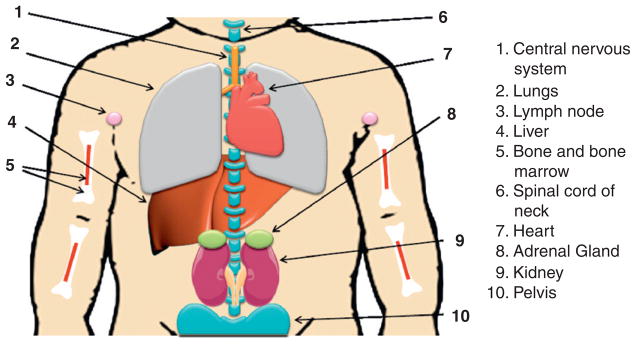
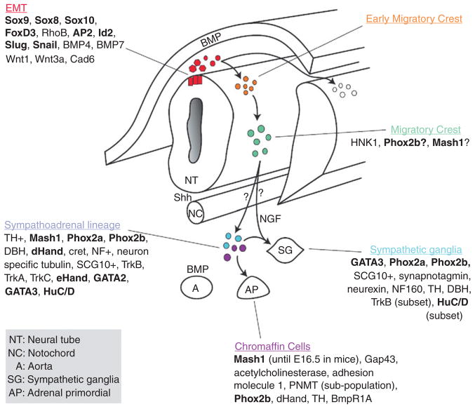
Neuroblastoma (NB), the most common extracranial solid tumor in childhood, is an extremely heterogeneous disease both biologically and clinically. Although significant progress has been made in identifying molecular and genetic markers for NB, this disease remains an enigmatic challenge. Since NB is thought to be an embryonal tumor that is derived from precursor cells of the peripheral (sympathetic) nervous system, understanding the development of normal sympathetic nervous system may highlight abnormal events that contribute to NB initiation. Therefore, this review focuses on the development of the peripheral trunk neural crest, the current understanding of how developmental factors may contribute to NB and on recent advances in the identification of important genetic lesions and signaling pathways involved in NB tumorigenesis and metastasis. Finally, we discuss how future advances in identification of molecular alterations in NB may lead to more effective, less toxic therapies, and improve the prognosis for NB patients.


Figures

Figure 4. 1
Figure 4. 1
Neuroblastoma localization. Neuroblastoma primary tumors derive from precursor cells of the peripheral (sympathetic) nervous system and can arise anywhere along the sympathetic chain, most frequently in the adrenal gland (position 8 as shown). Neuroblastoma may also develop from spinal cord of neck (position 6) and pelvis (position 10). Neuroblastomas mainly metastasize to lymph nodes (position 3), liver (position 4), bone and bone marrow (position 5), and also spread to central nervous system (position 1) and lungs (position 2) in infants.
Figure 4. 2
Figure 4. 2
General schema of the development of chromaffin cells and sympathetic ganglia. Cells at the dorsal region of the neural tube undergo EMT (red population), delaminate from the neural tube (orange), and migrate ventrally to the aorta (green) where they are commonly referred to as the sympathoadrenal progenitors (blue and purple). From the aortic region, the cells then migrate to the developing adrenal gland (AP) to become chromaffin cells or differentiate to become sympathetic ganglia (SG). As cells begin to differentiate as sympathetic ganglia they upregulate neural markers while chromaffin cells upregulate proteins found in the adrenal gland. Recent studies suggest that the chromaffin cell and sympathetic ganglia may come from divergent lineages rather than a common sympathoadrenal lineage. The question marks between the migrating crest and the sympathoadrenal progenitors address this possibility. A more detailed discussion on the temporal expression of the transcription factors in the sym-pathoadreanal lineage (including developmental stages) can be found in (Howard et al., 2000) and details about the distinct chromaffin and sympathetic lineages can be found in (Ernsberger et al., 2005). Additional of neurotrophins and their receptors can be found in (Straub et al., 2007). Transcription Factors are shown in bold and factors implicated in neuroblastoma have been underlined. Abbreviations: NT, Neural Tube; NC, notochord; A, Aorta; SG, sympathetic ganglia; AP, adrenal primordial.
Figure 4. 3
Figure 4. 3
The structure of N-myc protein and transcription regulation by N-myc. (A) The structure of N-myc protein. The N-terminal transactivation domain (TAD) contains two conserved Myc box I and II (MBI and MBII), which are essential for DNA binding. The C-terminal domain (CTD) harbors basic region (BR), helix-loop-helix (HLH) motif, and leucine zipper (LZ) for dimerization with Max. There is a nuclear localization signal (NLS) before CTD. (B) The model for the transcription regulation by N-myc. Myc–Max heterodimer may bind to E-box element (CACGTG) to activate transcription, however, Myc–Max dimer can associate with other transcription factors such as Miz-1, Smad, and bind to Inr (initiator, weak consensus) element to repress transcription. Max can also form homodimers or heterodimers with Mad to compete or suppress Myc–Max binding to E-box.

References

    1. Abe M, Ohira M, Kaneda A, Yagi Y, Yamamoto S, Kitano Y, Takato T, Nakagawara A, Ushijima T. CpG island methylator phenotype is a strong determinant of poor prognosis in neuroblastomas. Cancer Res. 2005;65:828–834. - PubMed
    1. Abe M, Westermann F, Nakagawara A, Takato T, Schwab M, Ushijima T. Marked and independent prognostic significance of the CpG island methylator phenotype in neuroblastomas. Cancer Lett. 2007;247:253–258. - PubMed
    1. Abel F, Sjoberg RM, Nilsson S, Kogner P, Martinsson T. Imbalance of the mitochondrial pro- and anti-apoptotic mediators in neuroblastoma tumours with unfavourable biology. Eur J Cancer. 2005;41:635–646. - PubMed
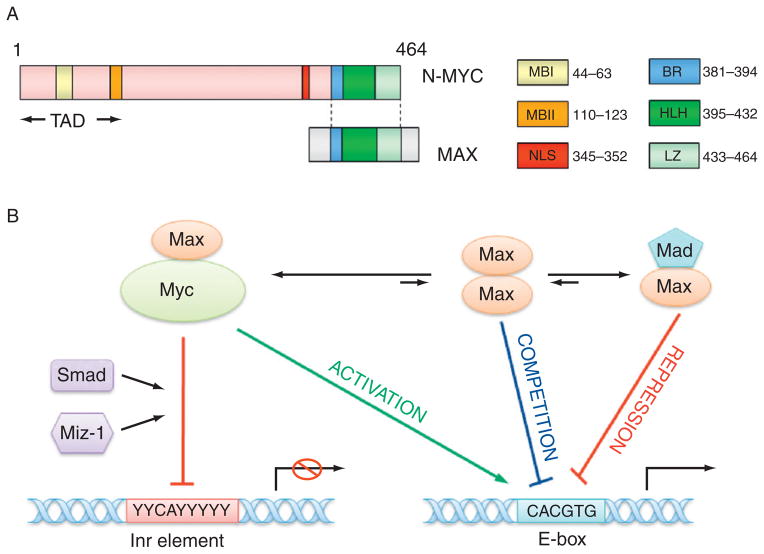
    1. Adhikary S, Eilers M. Transcriptional regulation and transformation by Myc proteins. Nat Rev Mol Cell Biol. 2005;6:635–645. - PubMed
    1. Alenina N, Bashammakh S, Bader M. Specification and differentiation of serotonergic neurons. Stem Cell Rev. 2006;2:5–10. - PubMed

Publication types

Substances