. 2011 May;32(13):3435-46.
doi: 10.1016/j.biomaterials.2011.01.021. Epub 2011 Feb 4.

The effect of surface charge on in vivo biodistribution of PEG-oligocholic acid based micellar nanoparticles

Affiliations

The effect of surface charge on in vivo biodistribution of PEG-oligocholic acid based micellar nanoparticles

Kai Xiao et al. Biomaterials. 2011 May.

Abstract

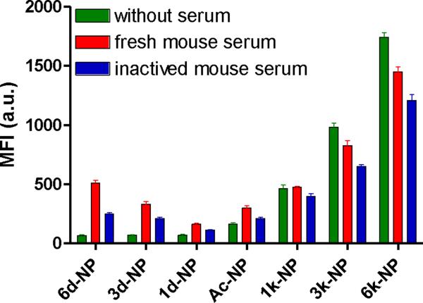
To systematically elucidate the effect of surface charge on the cellular uptake and in vivo fate of PEG-oligocholic acid based micellar nanoparticles (NPs), the distal PEG termini of monomeric PEG-oligocholic acid dendrimers (telodendrimers) are each derivatized with different number (n = 0, 1, 3 and 6) of anionic aspartic acids (negative charge) or cationic lysines (positive charge). Under aqueous condition, these telodendrimers self-assemble to form a series of micellar NPs with various surface charges, but with similar particle sizes. NPs with high surface charge, either positive or negative, were taken up more efficiently by RAW 264.7 murine macrophages after opsonization in fresh mouse serum. Mechanistic studies of cellular uptake of NPs indicated that several distinct endocytic pathways (e.g., clathrin-mediated endocytosis, caveolae-mediated endocytosis, and macropinocytosis) were involved in the cellular uptake process. After their cellular uptake, the majority of NPs were found to localize in the lysosome. Positively charged NPs exhibited dose-dependent hemolytic activities and cytotoxicities against RAW 264.7 cells proportional to the positive surface charge densities; whereas negatively charged NPs did not show obvious hemolytic and cytotoxic properties. In vivo biodistribution studies demonstrated that undesirable liver uptake was very high for highly positively or negatively charged NPs, which is likely due to active phagocytosis by macrophages (Kupffer cells) in the liver. In contrast, liver uptake was very low but tumor uptake was very high when the surface charge of NPs was slightly negative. Based on these studies, we can conclude that slightly negative charge may be introduced to the NPs surface to reduce the undesirable clearance by the reticuloendothelial system (RES) such as liver, improve the blood compatibility, thus deliver the anti-cancer drugs more efficiently to the tumor sites.


Figures

Fig.1
Fig.1
Schematic representation of the design of PEG5k-CA8 micellar NPs with various surface charges. Different number (n = 0, 1, 3 and 6) of D-aspartic acids (d) or D-lysines (k), which have anionic or cationic characters at physiological pH (7.4), respectively, were conjugated onto the distal end of PEG strands constituting a shell layer of the PEG5k-CA8 micellar NPs to modulate the surface charge. The N-terminal amino group was acetylated.
Fig.2
Fig.2
The morphology and particle size of different charged PEG5k-CA8 NPs measured by transmission electron microscopy (TEM).
Fig.3
Fig.3
Electrophoresis of DiD labeled PEG5k-CA8 NPs with various surface charge densities in 1% agarose gel. The DiD signals in the NPs were imaged using Kodak imaging station with excitation at 625 nm and emission at 700 nm.
Fig.4
Fig.4
Confocal fluorescent microscopy showing cellular uptake of NPs in RAW 264.7 murine macrophages after 2 hours of incubation with DiD-labeled PEG5k-CA8 NPs with various surface charges in serum-free medium.
Fig.5
Fig.5
The effect of the pre-opsonization of NPs in fresh or inactive mouse serum on their cellular uptake in RAW 264.7 murine macrophages. Data represent mean ± SEM (n = 3).
Fig.6
Fig.6
Intracellular tracking of different charged PEG5k-CA8 NPs in RAW 264.7 macrophages. RAW 264.7 macrophages were incubated with DiD (red) labeled PEG5k-CA8 NPs with various surface charges for 2 hours in medium with 10% fresh mouse serum, after which the cells were labeled with lysosome tracker (green) for 30 min before imaging by confocal microscopy. Co-localization of NPs with lysosome appears yellow in merged images, indicating that the NPs are present in the lysosome.
Fig.7
Fig.7
The cellular uptake pathways of different charged PEG5k-CA8 NPs in macrophages. RAW 264.7 cells were either pre-incubated at 4 °C for 3 h, followed by incubation with DiD-labeled PEG5k-CA8 NPs with various surface charges at 4 °C for 2 h; or pre-treated with different endocytosis inhibitors such as filipin III (1 μg/mL), amiloride (50 μM) and chlorpromazine (CPZ, 10 μg/mL) at 37 °C for 1 h, followed by incubation with different charged PEG5k-CA8 NPs at 37 °C for 2 h. (A) Confocal microscopy images of cellular uptake. Nuclei were stained by DAPI (blue). (B) Quantitative analysis of the cellular uptake by flow cytometry. Data represent mean ± SEM (n = 3).
Fig.8
Fig.8
In vitro red blood cells (RBCs) lysis. Different charged PEG5k-CA8 NPs were incubated with human erythrocyte suspension for 4 h at 37 °C. RBCs lysis was determined spectrophotometrically (λ = 540 nm) based on hemoglobin level. PBS was used as negative control, and Triton-100 was used as positive control. Data represent mean ± SEM (n = 3).
Fig.9
Fig.9
The effect of surface charge on the cell viability of PEG5k-CA8 NPs against RAW 264.7 cells measured by MTT assay. Data represent mean ± SEM (n = 3). *: p < 0.05, **: p < 0.01, ***: p < 0.001.
Fig.10
Fig.10
Biodistribution of different charged PEG5k-CA8 NPs after intravenous injection in SKOV-3 tumor bearing mice. (A) Representative ex vivo near-infrared (NIRF) optical images of tumor and major organs excised at 24 h after intravenous injection of DiD loaded PEG5k-CA8 NPs with various surface charges. (B) Quantitative fluorescence intensities of tumors and organs from ex vivo images (n = 3). (C) Microscopic images of liver cryo-section. The nuclei were stained by DAPI (blue), and macrophages were stained by F4/80 antibody (yellow), and the signals of DiD (red) were acquired using Olympus FV1000 laser scanning confocal microscopy. *: p < 0.05, **: p < 0.01, ***: p < 0.001.

Similar articles

Cited by

References

    1. Matsumura Y, Maeda H. A new concept for macromolecular therapeutics in cancer chemotherapy: mechanism of tumoritropic accumulation of proteins and the antitumor agent smancs. Cancer Res. 1986;46(12 Pt 1):6387–92. - PubMed
    1. Alexis F, Pridgen E, Molnar LK, Farokhzad OC. Factors affecting the clearance and biodistribution of polymeric nanoparticles. Mol Pharm. 2008;5(4):505–15. - PMC - PubMed
    1. Davis ME, Chen ZG, Shin DM. Nanoparticle therapeutics: an emerging treatment modality for cancer. Nat Rev Drug Discov. 2008;7(9):771–82. - PubMed
    1. Gref R, Minamitake Y, Peracchia MT, Trubetskoy V, Torchilin V, Langer R. Biodegradable long-circulating polymeric nanospheres. Science. 1994;263(5153):1600–3. - PubMed
    1. Zahr AS, Davis CA, Pishko MV. Macrophage uptake of core-shell nanoparticles surface modified with poly(ethylene glycol) Langmuir. 2006;22(19):8178–85. - PubMed

Publication types