

Placental volume and vascular flow assessed by 3D power Doppler and adverse pregnancy outcomes
- PMID: 21295850
- PMCID: PMC3125967
- DOI: 10.1016/j.placenta.2011.01.010
Placental volume and vascular flow assessed by 3D power Doppler and adverse pregnancy outcomes
Abstract
Objective: To estimate the utility of first-trimester 3D-placental volume and vascular flow indices in the prediction of adverse pregnancy outcomes.
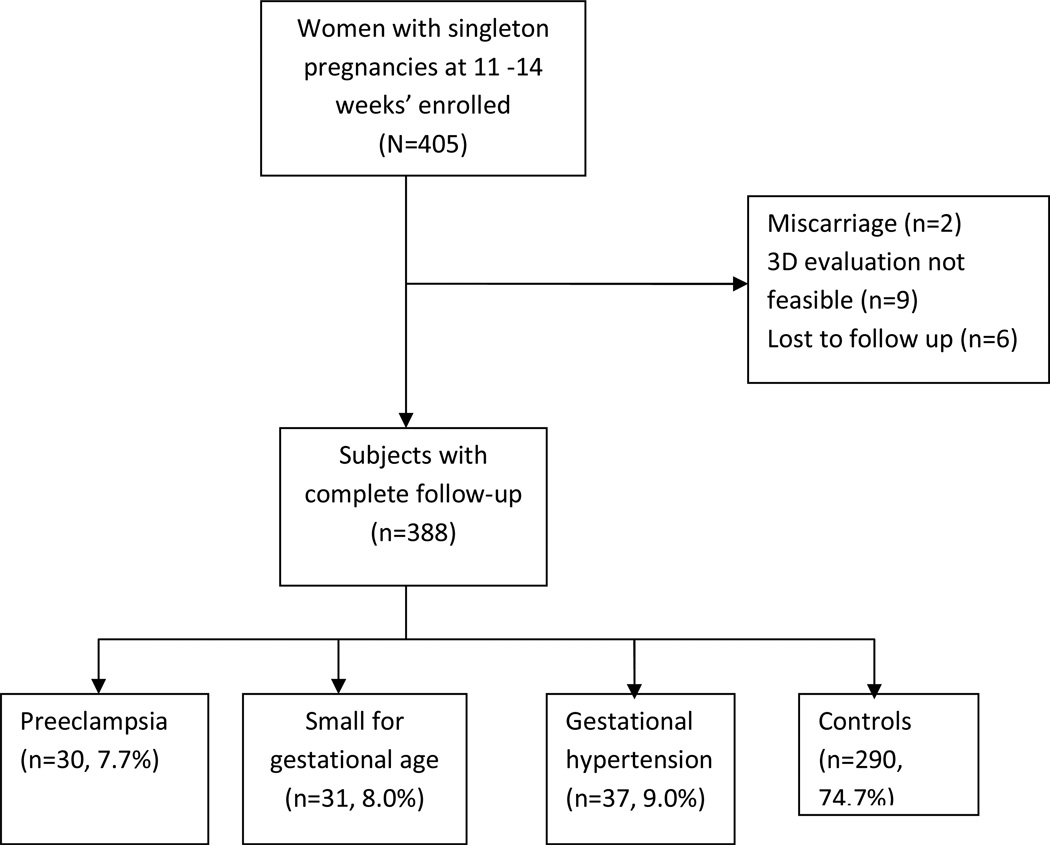
Methods: A prospective cohort study including women with singleton pregnancies seen between 11 and 14 weeks as part of a screening program for aneuploidy. Placental volume and vascularization indices were obtained using 3D power Doppler imaging and the VOCAL technique. Placental volume (PV), Vascularization Index (VI), Flow Index (FI) and Vascularization Flow Index (VFI) were calculated. The adverse pregnancy outcomes investigated include preeclampsia (PE), gestational hypertension (GH) and small for gestational age (SGA). The predictive ability of each variable was evaluated using receiver-operating characteristic (ROC) curves.
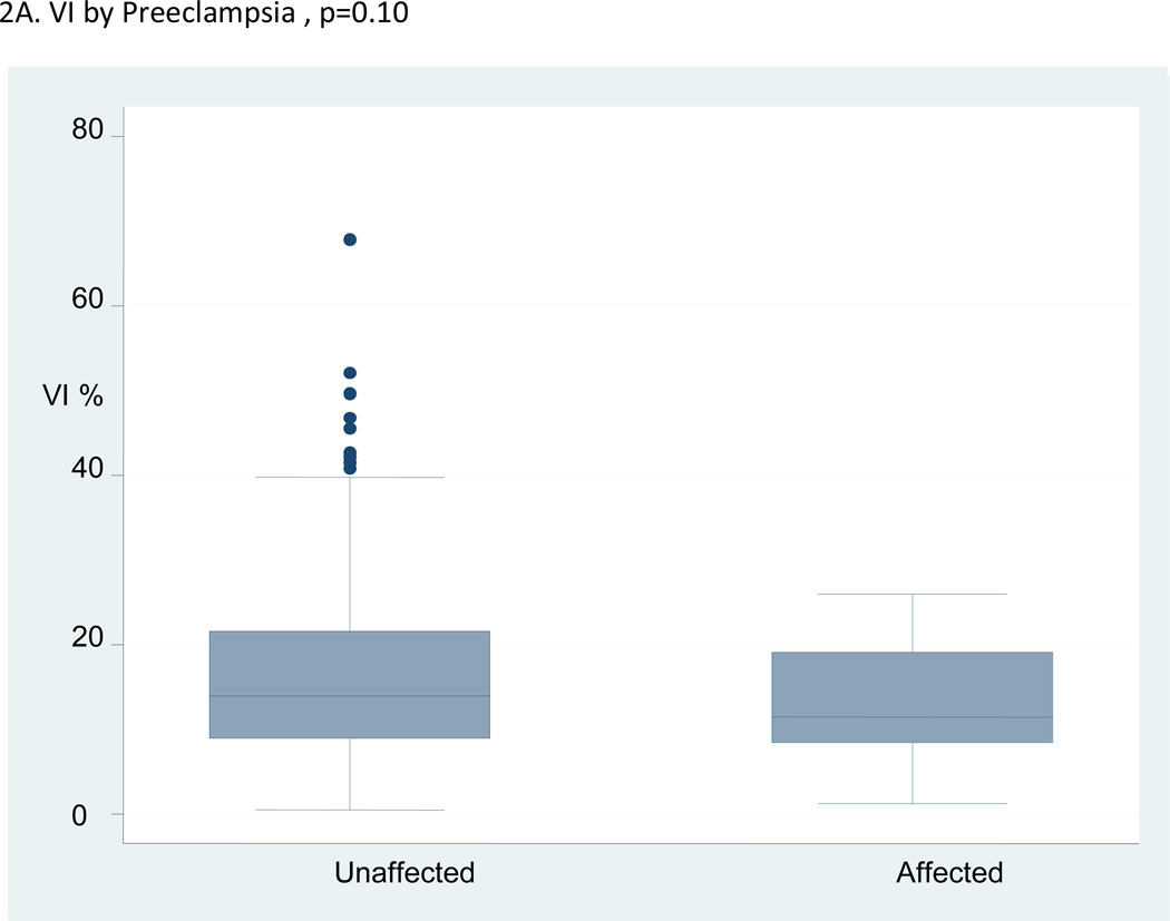
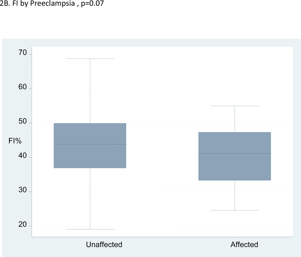
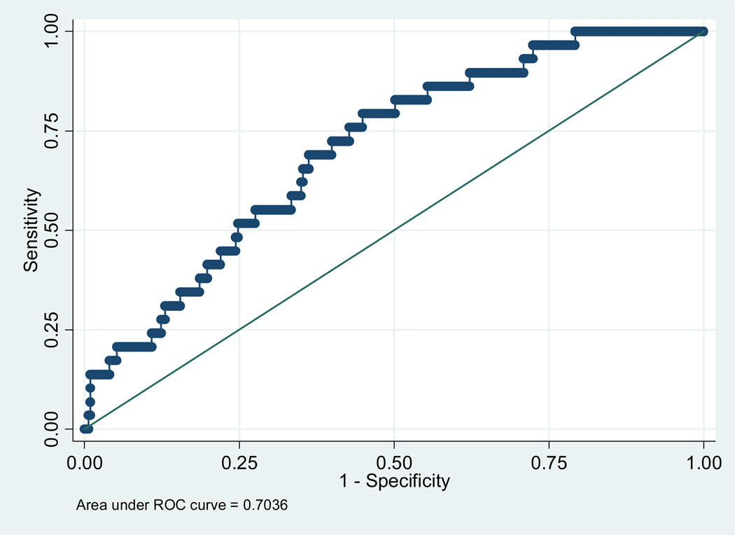
Results: Of 388 women included, PE was seen in 30 (7.7%), GH in 37 (9.0%) and SGA in 31 (8.0%). Placental volume was not significantly different between the pregnancies with adverse outcomes and those without. The mean values of the VI and VFI were significantly lower in the pregnancies that developed PE but not in GH or SGA. The area under the ROC curve for the prediction of PE was 0.71, 0.69 and 0.70 for VI, FI and VFI, respectively.
Conclusion: The study confirms lower 3D power Doppler vascular flow indices in pregnancies that develop PE. The discriminatory ability of using these indices alone for predicting PE appears modest.
Copyright © 2011 Elsevier Ltd. All rights reserved.
Figures

References
-
- Hafner E, Philipp T, Schuchter K, Dillinger-Paller B, Philipp K, Bauer P. Second-trimester measurements of placental volume by three-dimensional ultrasound to predict small-for gestational- age infants. Ultrasound Obstet Gynecol. 1998;12:97–102. - PubMed
-
- Pairleitner H, Steiner H, Hasenoehrl G, Staudach A. Three-dimensional power Doppler sonography: imaging and quantifying blood flow and vascularization. Ultrasound Obstet Gynecol. 1999;14:139–143. - PubMed
-
- Hafner T, Kurjak A, Funduk-Kurjak B, Bekavac I. Assessment of early chorionic circulation by three-dimensional power Doppler. J Perinat Med. 2002;30(1):33–39. - PubMed
-
- Matijevic R, Kurjak A. The assessment of placental blood vessels by three-dimensional power Doppler ultrasound. J Perinat Med. 2002;30:26–32. - PubMed
-
- Pretorius DH, Nelson TR, Baergen RN, Pai E, Cantrell C. Imaging of placental vasculature using three-dimensional ultrasound and color power Doppler: a preliminary study. Ultrasound Obstet Gynecol. 1998;12:45–49. - PubMed
MeSH terms
Grants and funding
LinkOut - more resources
Full Text Sources
