Review
. 2011 Mar 31;367(1-2):1-16.
doi: 10.1016/j.jim.2011.01.015. Epub 2011 Feb 4.

Methodology and application of flow cytometry for investigation of human malaria parasites

Affiliations
Review

Methodology and application of flow cytometry for investigation of human malaria parasites

Brian T Grimberg. J Immunol Methods. .

Abstract

Historically, examinations of the inhibition of malaria parasite growth/invasion, whether using drugs or antibodies, have relied on the use of microscopy or radioactive hypoxanthine uptake. These are considered gold standards for measuring the effectiveness of antimalarial treatments, however, these methods have well known shortcomings. With the advent of flow cytometry coupled with the use of fluorescent DNA stains allowed for increased speed, reproducibility, and qualitative estimates of the effectiveness of antibodies and drugs to limit malaria parasite growth which addresses the challenges of traditional techniques. Because materials and machines available to research facilities are so varied, different methods have been developed to investigate malaria parasites by flow cytometry. This review is intended to serve as a reference guide for advanced users and importantly, as a primer for new users, to support expanded use and improvements to malaria flow cytometry, particularly in endemic countries.


Figures

Figure 1
Figure 1
History of Flow Cytometry and Analysis of Human Malaria Parasites
Figure 2
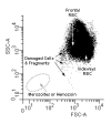
Figure 2. Identification of Populations from Forward Scatter/Side Scatter of Red Blood Cells
The biconcave shape of the red blood cell allows for the cell to pass through the flow core of the cytometer with the lasers passing through its front in the center of the cell which is 2 microns thick or pass through sideways where the laser passes through its’ width which is 6 microns across. This accounts for the two major populations observed. In any given sample their can also be platelets or cell fragments which can confound further analysis of the malaria parasites. Also a combination of hemozoin (of varying sizes) and free merozoites can be found in some samples. The hemozoin populations can be removed by the magnetic separation and the merozoites are positive for the presence of 1N copy of DNA (Boyle et al., 2010).
Figure 3
Figure 3. Comparisons of Different DNA stains
A sample of P.falciparum (3D7 Strain, MRA-102 deposited to ATCC/MR4 by DJ Carucci) was cultured as previously indicated (McNamara et al., 2006) to a parasitemia of 2.9% as determined by Giemsa stained microscopy slide. This culture was then exposed to stains commonly used to examine malaria. Though there may be more optimal methods, staining was carried out as indicated in references; Ethidium Bromide (Persson et al., 2006), Propidium Iodide (Pattanapanyasat et al., 1997), Thiazole Orange (Makler et al., 1987), SYBR Green (Dent et al., 2008), YOYO-1 (Dahl and Rosenthal, 2007), Hoechst 33342 (Grimberg et al., 2008) and the resulting frequency of positive cells is indicated. The blood sample used for this study were leukocyte depleted using Histopaque 1119 and had been in culture for 3 days and therefore, there were few reticulocytes left at the time of analysis. In addition, the parasites had been serial transferred two times after thawing from frozen stocks which led to very clean samples, (i.e. containing very few dead parasites or cell fragments), which led to 5 out of the 6 stains tested showing the same parasitemia. When dealing with samples from patients all of the above issues can confuse a clear delineation between uninfected and infected cells. This figure shows that when using a well prepared in vitro sample with a moderate parasite load, many of the commonly used stains can determine parasitemia.
Figure 4
Figure 4. Identification of Erythrocyte Lifecycle Stages of P. falciparum
The same sample of 2.9% parasitemia culture of P. falciparum used in Figure 3 was stained with HO342, TO, and DiIC1-5 as described in Grimberg et al. (2009). Gates positions were determined previously (Grimberg et al., 2008; Grimberg et al., 2009). In brief, DNA positive cells (Y-axis, UV 440, HO342) in the left hand panel which showed membrane potential (X axis, Red 660, DiIC1-5) greater than that of the red blood cells were considered to be alive. The slanted area of this gate was determined by the application of a cyanide derivative (carbonyl cyanide 3-chlorophenylhydrazone) which disrupts membrane potential within the cells. In the right hand panel, the DNA negative uninfected cells (from the gray square) are shown along with the Live DNA positive cells (black polygon). By showing the relative amount of RNA in these cells one can clearly identify the different erythrocyte life cycle stages of the parasites. Ring stages do not express very much RNA, trophozoites express RNA but do not replicate DNA, and schizonts have increases in both RNA and DNA. The delineation between schizonts and trophozoites has been made between 3 and 4 copies of DNA (Grimberg et al., 2008) in general because of the lack of 4N rings (therefore 4N DNA/RNA positive cells almost exclusively arise through DNA synthesis).

Similar articles

Cited by

References

    1. Adams JH, Sim BK, Dolan SA, Fang X, Kaslow DC, Miller LH. A family of erythrocyte binding proteins of malaria parasites. Proc Natl Acad Sci USA. 1992;89:7085–9. - PMC - PubMed
    1. Ahlborg N, Iqbal J, Bjork L, Stahl S, Perlmann P, Berzins K. Plasmodium falciparum: differential parasite growth inhibition mediated by antibodies to the antigens Pf332 and Pf155/RESA. Exp Parasitol. 1996;82:155–63. - PubMed
    1. Angulo-Barturen I, Jimenez-Diaz MB, Mulet T, Rullas J, Herreros E, Ferrer S, Jimenez E, Mendoza A, Regadera J, Rosenthal PJ, Bathurst I, Pompliano DL, de las Heras F. Gomez, Gargallo-Viola D. A murine model of falciparum-malaria by in vivo selection of competent strains in non-myelodepleted mice engrafted with human erythrocytes. PLoS One. 2008;3:e2252. - PMC - PubMed
    1. Arndt-Jovin DJ, Jovin TM. Analysis and sorting of living cells according to deoxyribonucleic acid content. J Histochem Cytochem. 1977;25:585–9. - PubMed
    1. Baniecki ML, Wirth DF, Clardy J. High-throughput Plasmodium falciparum growth assay for malaria drug discovery. Antimicrob Agents Chemother. 2007;51:716–23. - PMC - PubMed

Publication types

LinkOut - more resources