

Bupivacaine decreases cell viability and matrix protein synthesis in an intervertebral disc organ model system
- PMID: 21296298
- PMCID: PMC3056334
- DOI: 10.1016/j.spinee.2010.11.017
Bupivacaine decreases cell viability and matrix protein synthesis in an intervertebral disc organ model system
Abstract
Background context: Bupivacaine is a local anesthetic commonly used for back pain management in interventional procedures. Cytotoxic effects of bupivacaine have been reported in articular cartilage and, recently, in intervertebral disc cell culture. However, the relevance of these effects to discs in vivo remains unclear. This study examines the effect of bupivacaine on disc cell metabolism using an organotypic culture model system that mimics the in vivo environment.
Purpose: To assess the effect of bupivacaine on disc cell viability and matrix protein synthesis using an organotypic model system and to determine whether this anesthetic has toxic effects.
Study design: Mouse intervertebral discs were isolated and maintained ex vivo in an organotypic culture then exposed to clinically relevant concentrations of bupivacaine, and the impact on disc cell viability and matrix proteoglycan (PG) and collagen syntheses were measured in the presence and absence of the drug.
Subjects: Mouse functional spine units (FSUs) were isolated from the lumbar spines of 10-week-old mice.
Outcome measures: Cell viability was assessed by 3-(4,5-dimethylthiazol-2-yl)-2,5-diphenyltetrazolium bromide (MTT) assay. Total PG and collagen syntheses were determined by measuring the incorporation of radioactive (35)S-sulfate and (3)H-l-proline into PG and collagen, respectively.
Methods: Organotypic cultures of mouse FSUs were exposed to different concentrations (0%-0.5%) of bupivacaine for variable amounts of time (0-2 hours). Cell viability within disc tissue was quantified by MTT staining and histologic assay. Matrix protein synthesis was measured by incorporation of radioactive (35)S-sulfate (for PG synthesis) and (3)H-l-proline (for collagen synthesis).
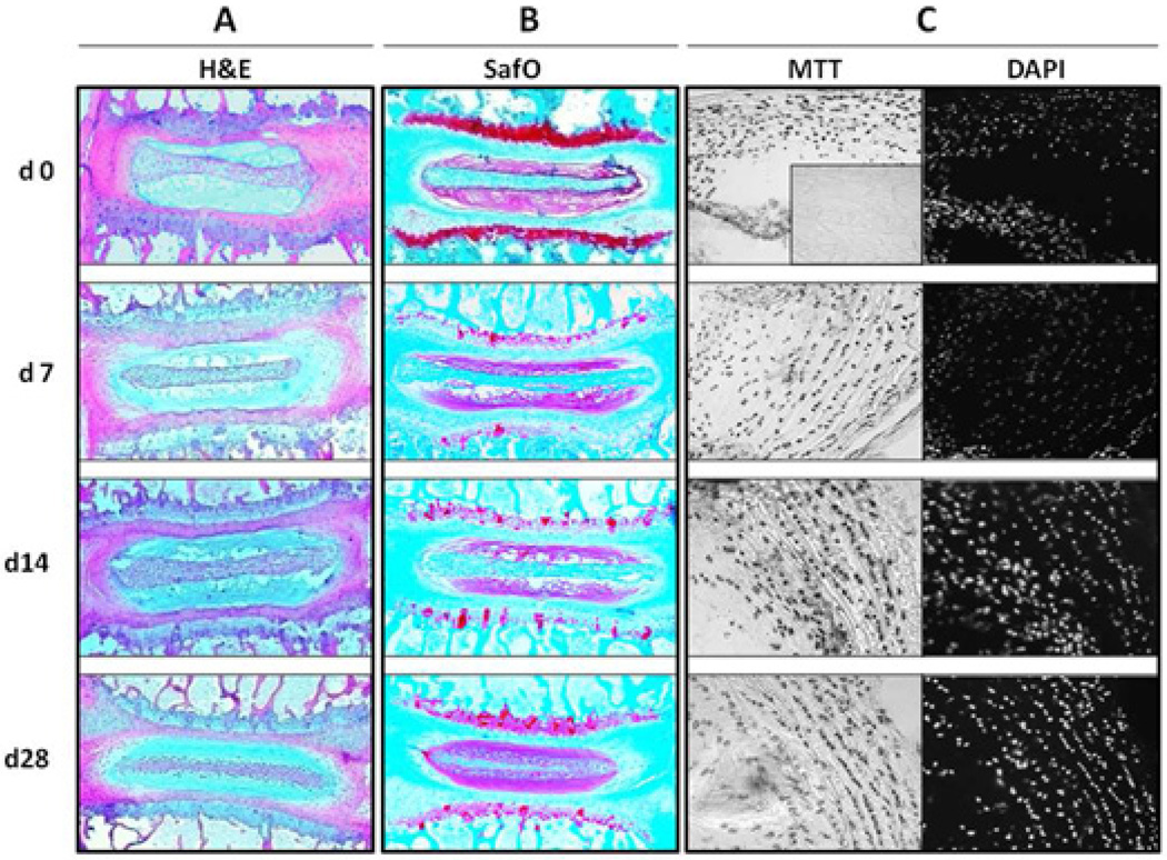
Results: Untreated mouse disc organs were maintained in culture for up to 1 month with minimal changes in tissue histology, cell viability, and matrix protein synthesis. Exposure to bupivacaine decreased cell viability in a dose- and time-dependent manner. Exposure to bupivacaine at concentrations less than or equal to 0.25% did not significantly affect matrix protein synthesis. However, at 0.5% bupivacaine, collagen synthesis was reduced by fourfold and PG synthesis by threefold.
Conclusions: Mouse discs can be successfully maintained ex vivo for upward of 4 weeks with little cell death, change in histologic structure, or matrix protein synthesis. This organotypic model system closely mimics the in vivo environment of the disc. Exposure of these cultures to bupivacaine dramatically decreased cell viability and matrix protein synthesis in a dose- and time-dependent manner. These findings corroborate those previously reported by Lee et al. using disc cell culture and demonstrate that this anesthetic at clinically relevant doses is toxic to intervertebral discs in both cell culture and disc organ models representative of the native architectural context.
Published by Elsevier Inc.
Figures


References
-
- Hart LG, Deyo RA, Cherkin DC. Physician office visits for low back pain. Frequency, clinical evaluation, and treatment patterns from a U.S. national survey. Spine. 1995;20:11–19. - PubMed
-
- Smith MD, McGhan WF. Treating back pain without breaking the bank. Bus Health. 1998;16:50–51. - PubMed
-
- Kelsey JL, White AA. Epidemiology and impact of low back pain. Spine. 1980;5:133–142. - PubMed
-
- Luo X, pietrobon R, Sun SX, Liu GG, Hey L. Estimates and patterns of direct health care expenditures among individuals with back pain in the United States. Spine. 2004;29:79–86. - PubMed
Publication types
MeSH terms
Substances
Grants and funding
LinkOut - more resources
Full Text Sources
