Review
. 2011 Jun;23(3):293-301.
doi: 10.1016/j.ceb.2011.01.002. Epub 2011 Feb 4.

Nuclear pore complex-a coat specifically tailored for the nuclear envelope

Affiliations
Review

Nuclear pore complex-a coat specifically tailored for the nuclear envelope

Evgeny Onischenko et al. Curr Opin Cell Biol. 2011 Jun.

Abstract

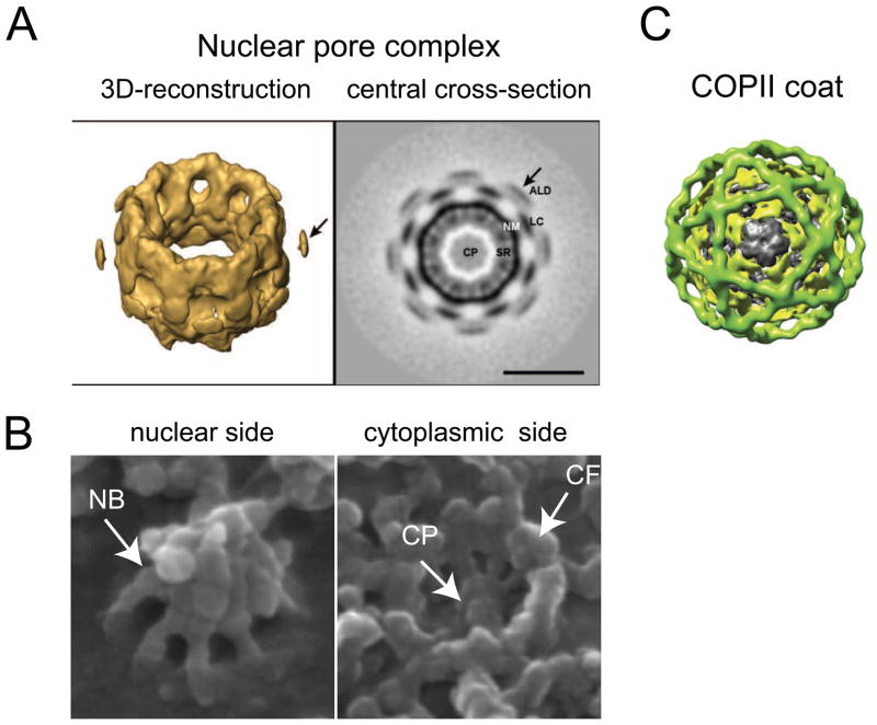
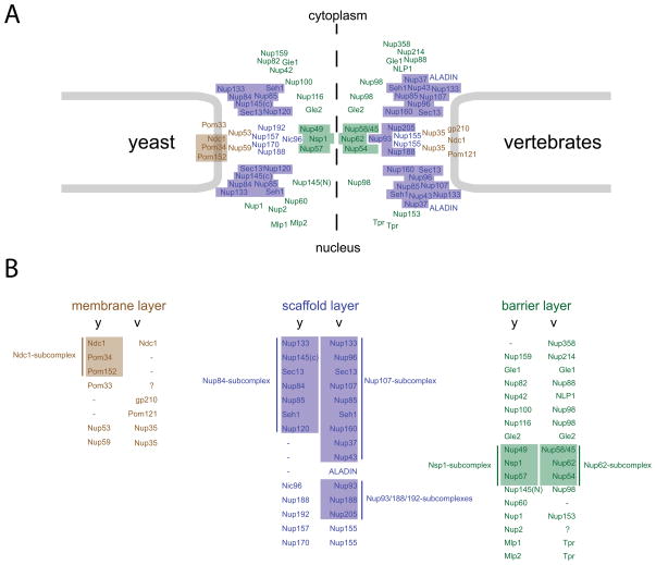
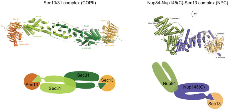
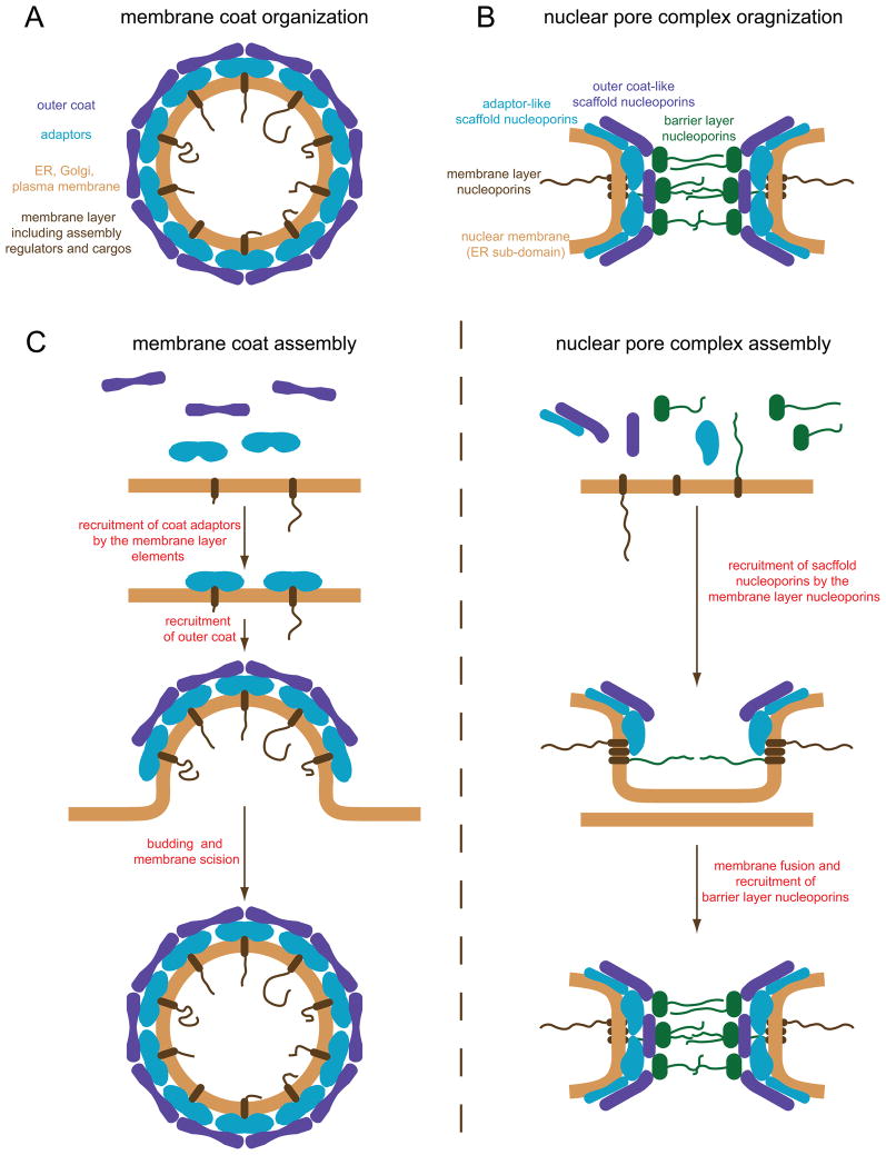
Nuclear pore complexes (NPCs) are highly selective transport gates that enable the bi-directional traffic of macromolecules across the nuclear envelope (NE). NPCs are located at the fusion pores between the inner and outer membranes of the NE and are built from a common set of ∼30 different proteins, nucleoporins. Remarkably, recent proteomic, bioinformatic, and structural studies have provided firm evidence that key structural nucleoporins share common ancestry with elements of coated vesicles, indicating an evolutionary link between these structures. This has provided novel insight into the origin of NPCs and may help us to better functionally characterize these fundamental components of eukaryotic cells.


Figures

Figure 1
Figure 1
Structural highlights of NPCs and COP/clathrin coats. (A) A 3D-reconstruction (left) or a central cross-section (right) of the NPC structure acquired by cryo-EM tomography using Xenopus NPCs. Note the characteristic 8-fold symmetry of the NPC. The image is adapted from [10•]. CP, central plug; SR, spoke ring complex; NM, nuclear membrane; LC, luminal connection; ALD, additional luminal density. (B) Surface views of the nuclear (left) and the cytoplasmic sides (right) of Xenopus NPC acquired using high-resolution scanning EM. The images highlight the nuclear basket (NB), cytoplasmic filaments (CF) and the central plug (CP). These structures are not clearly visible with cryo-EM tomography (Fig. 1A) due to their dynamic nature. Images were kindly provided by Dr. Elena Kiseleva. (C) Architecture of the COPII coat visualized by EM single particle reconstruction. Note the spherical architecture and highly symmetrical arrangement of coatomers within the coat structure (adapted from [30]).
Figure 2
Figure 2
NPC composition in yeast and vertebrates. (A) Panel displays the distribution and approximate location of yeast nucleoporins (left) and corresponding vertebrate nucleoporins (right) within the NPC. Based on their functional properties and localization nucleoporins can be classified into 3 functional layers: the membrane layer (brown) consisting of transmembrane and membrane-associated nucleoporins, the scaffold layer (blue) composed of core structural nucleoporins, and the barrier layer (green) containing nucleoporins involved in the selective NPC permeability and other functions. (B) The correspondence between yeast (y) and vertebrate (v) nucleoporins. Colored boxes depict biochemically stable nucleoporin subcomplexes.
Figure 3
Figure 3
Structural similarities between COPII coat elements and scaffold nucleoporins as exemplified by the Nup84-Nup145(C)-Sec13 complex structure. The top panels show structures of the Sec13/31 COPII coatomer complex (left) and the Nup84-Nup145(C)-Sec13 nucleoporin complex (right). The bottom panels display schematic representations of these complexes highlighting (a) the characteristic U-shaped organization of Nup84 and Nup145(C), which is similar to the arrangement of the Sec31 alpha-helical domain, (b) the anti-parallel mode of interaction between Nup84 and Nup145(C) and two Sec31 molecules, and (c) the in-trans blade insertions of Nup145(C) or Sec31 into the Sec13 beta-propeller. For a detailed description of these similarities see [19••,23] and main text. The structures were adapted from [23,26].
Figure 4
Figure 4
(A and B) Models of the membrane coat (A) and NPC organization (B). Functionally similar parts of NPCs and membrane coats are shown in the same color. (C) Corresponding steps in the assembly of membrane coats (left) and NPCs (right).

References

    1. Neumann N, Lundin D, Poole AM. Comparative genomic evidence for a complete nuclear pore complex in the last eukaryotic common ancestor. PLoS One. 2010;5:e13241. - PMC - PubMed
    1. Mans BJ, Anantharaman V, Aravind L, Koonin EV. Comparative genomics, evolution and origins of the nuclear envelope and nuclear pore complex. Cell Cycle. 2004;3:1612–1637. - PubMed
    1. Rout MP, Aitchison JD, Suprapto A, Hjertaas K, Zhao Y, Chait BT. The yeast nuclear pore complex: composition, architecture, and transport mechanism. J Cell Biol. 2000;148:635–51. - PMC - PubMed
    1. DeGrasse JA, DuBois KN, Devos D, Siegel TN, Sali A, Field MC, Rout MP, Chait BT. Evidence for a shared nuclear pore complex architecture that is conserved from the last common eukaryotic ancestor. Mol Cell Proteomics. 2009;8:2119–2130. This paper describes the proteomic analysis of NPCs from Trypanosoma brucei, a highly divergent eukaryote, evolutionarily distant from species whose NPCs have been analyzed previously. The authors find that in spite of low sequence homology the majority of experimentally identified nucleoporins in trypanosomes have clear orthologs in yeast and vertebrates. Intriguingly, NPCs from T. brucei contain both scaffold and barrier layer nucleoporins, but transmbrane nucleoporins were not identified. Together these results suggest that a common NPC core structure was already present in the last eukaryotic common ancestor. - PMC - PubMed
    1. Cronshaw JM, Krutchinsky AN, Zhang W, Chait BT, Matunis MJ. Proteomic analysis of the mammalian nuclear pore complex. J Cell Biol. 2002;158:915–27. - PMC - PubMed

Publication types

Substances