

Effect of topical azithromycin on corneal innate immune responses
- PMID: 21296809
- PMCID: PMC3262402
- DOI: 10.1167/iovs.10-5658
Effect of topical azithromycin on corneal innate immune responses
Abstract
Purpose: To determine the effect of azithromycin (AZM) in a murine model of corneal inflammation.
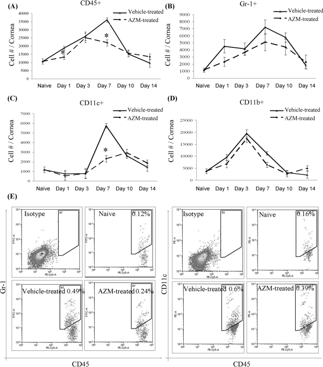
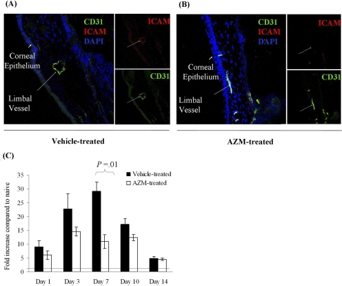
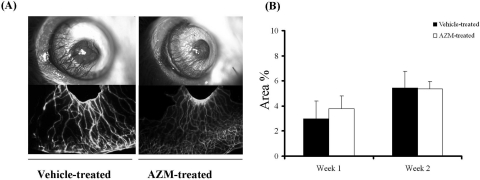
Methods: The effect of topical AZM was studied in murine corneal inflammation. Corneal inflammation was induced by thermal cautery in BALB/c mice. Leukocyte infiltration at different time points was analyzed by flow cytometry. At set time points, real-time polymerase chain reaction was performed to quantify the expression of different inflammatory cytokine transcript in the cornea. Corneal samples were analyzed immunohistochemically for the expression of intercellular adhesion molecule-1 (ICAM-1). Corneal neovascularization (CNV) was induced by micropellet (VEGF-A) placement. Mice were then treated topically with either AZM or vehicle. CNV was evaluated morphometrically.
Results: Eyes receiving AZM showed a significant decrease in corneal infiltration compared with the vehicle-treated group. AZM also significantly decreased messenger RNA expression levels of interleukin-1β (IL-1β), tumor necrosis factor-α (TNF-α) and ICAM-1 in the cornea. There was no significant difference in CNV between the AZM- and vehicle-treated groups.
Conclusions: After an inflammatory insult, topical AZM significantly reduced leukocyte infiltration into the cornea. This was further supported by an associated decrease in expression of IL-1β, TNF-α, and ICAM-1 in the cornea, indicating AZM may have a potential anti-inflammatory effect on corneal inflammation.
Figures

References
-
- Streilein JW. Immunology and immunopathology of corneal transplantation. Chem Immunol. 1999;73:186–206 - PubMed
-
- Dana MR. Corneal antigen-presenting cells: diversity, plasticity, and disguise: the Cogan lecture. Invest Ophthalmol Vis Sci. 2004;45:722–727 - PubMed
-
- Delves PJ, Roitt IM. The immune system: first of two parts. N Engl J Med. 2000. 6;343:37–49 - PubMed
-
- Hamrah P, Huq SO, Liu Y, Zhang Q, Dana MR. Corneal immunity is mediated by heterogeneous population of antigen presenting cells. J Leukoc Biol. 2003;74:172–178 - PubMed
-
- Hamrah P, Zhang Q, Liu Y, Dana MR. Novel characterization of MHC class II–negative population of resident corneal Langerhans cell–type dendritic cells. Invest Ophthalmol Vis Sci. 2003;43:639–646 - PubMed
Publication types
MeSH terms
Substances
Grants and funding
LinkOut - more resources
Full Text Sources
Medical
Miscellaneous
