

The impact of retardance pattern variability on nerve fiber layer measurements over time using GDx with variable and enhanced corneal compensation
- PMID: 21296821
- PMCID: PMC3175933
- DOI: 10.1167/iovs.10-5969
The impact of retardance pattern variability on nerve fiber layer measurements over time using GDx with variable and enhanced corneal compensation
Abstract
Purpose: To examine the impact of retardance pattern variability on retinal nerve fiber layer (RNFL) measurements over time using scanning laser polarimetry with variable (GDxVCC) and enhanced corneal compensation (GDxECC; both by Carl Zeiss Meditec, Inc., Dublin, CA).
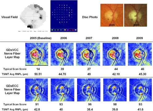
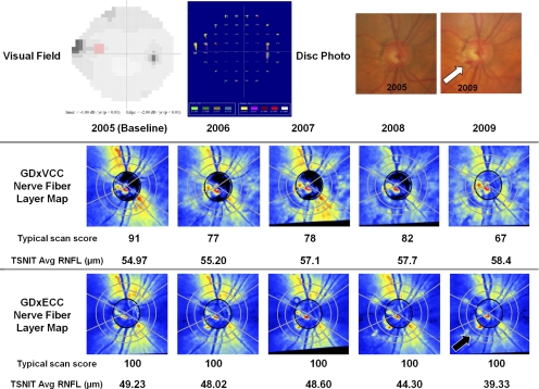
Methods: Glaucoma suspect and glaucomatous eyes with 4 years of follow-up participating in the Advanced Imaging in Glaucoma Study were prospectively enrolled. All eyes underwent standard automated perimetry (SAP), GDxVCC, and GDxECC imaging every 6 months. SAP progression was determined with point-wise linear regression analysis of SAP sensitivity values. Typical scan score (TSS) values were extracted as a measure of retardance image quality; an atypical retardation pattern (ARP) was defined as TSS < 80. TSS fluctuation over time was measured using three parameters: change in TSS from baseline, absolute difference (maximum minus minimum TSS value), and TSS variance. Linear mixed-effects models that accommodated the association between the two eyes were constructed to evaluate the relationship between change in TSS and RNFL thickness over time.
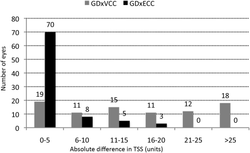
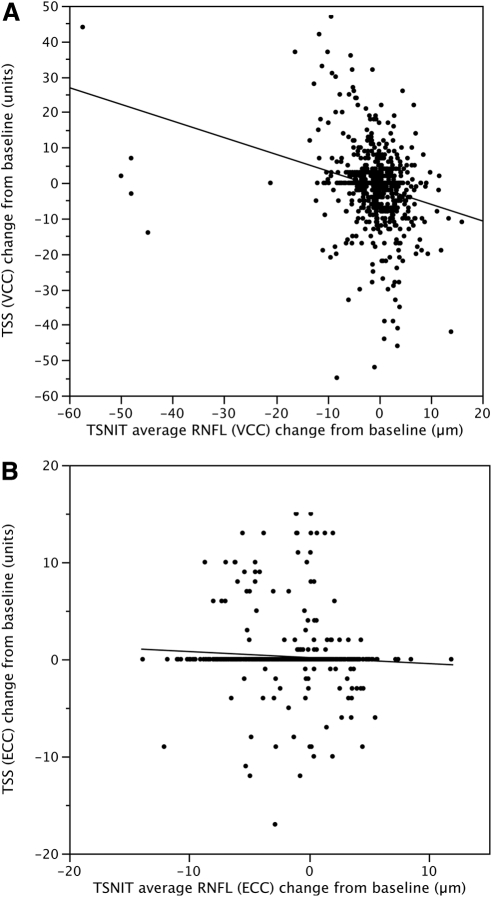
Results: Eighty-six eyes (51 suspected glaucoma, 35 glaucomatous) of 45 patients were enrolled. Twenty (23.3%) eyes demonstrated SAP progression. There was significantly greater fluctuation in TSS over time with GDxVCC compared with GDxECC as measured by absolute difference (18.40 ± 15.35 units vs. 2.50 ± 4.69 units; P < 0.001), TSS variance (59.63 ± 87.27 units vs. 3.82 ± 9.63 units, P < 0.001), and change in TSS from baseline (-0.83 ± 11.2 vs. 0.25 ± 2.9, P = 0.01). The change in TSS over time significantly (P = 0.006) influenced the TSNIT average RNFL thickness when measured by GDxVCC but not by GDxECC.
Conclusions: Longitudinal images obtained with GDxECC have significantly less variability in TSS and retardance patterns and have reduced bias produced by ARP on RNFL progression assessment.
Figures
References
-
- Greenfield DS, Knighton RW, Huang XR. Effect of corneal polarization axis on assessment of retinal nerve fiber layer thickness by scanning laser polarimetry. Am J Ophthalmol. 2000;129:715–722 - PubMed
-
- Knighton RW, Huang XR, Greenfield DS. Analytical model of scanning laser polarimetry for retinal nerve fiber layer assessment. Invest Ophthalmol Vis Sci. 2002;43:383–392 - PubMed
-
- Knighton RW, Huang X, Zhou Q. Microtubule contribution to the reflectance of the retinal nerve fiber layer. Invest Ophthalmol Vis Sci. 1998;39:189–193 - PubMed
-
- Weinreb RN, Bowd C, Greenfield DS, Zangwill LM. Measurement of the magnitude and axis of corneal polarization with scanning laser polarimetry. Arch Ophthalmol. 2002;120:901–906 - PubMed
-
- Weinreb RN, Bowd C, Zangwill LM. Glaucoma detection using scanning laser polarimetry with variable corneal polarization compensation. Arch Ophthalmol. 2003;121:218–224 - PubMed
Publication types
MeSH terms
Grants and funding
LinkOut - more resources
Full Text Sources
Medical
Miscellaneous
